
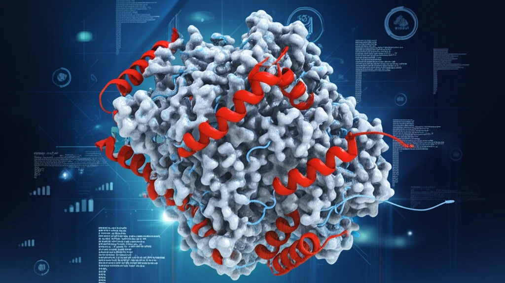
Decoding Protein Structures: A Guide to Model Evaluation

"Navigating the complexities of protein analysis for groundbreaking advancements in biomedicine and beyond."
Proteins are the workhorses of our cells, and understanding their structure is essential for understanding their function. This knowledge is the bedrock of advancements in biomedicine, drug discovery, and biotechnology. The ability to accurately predict and model protein structures is thus a critical pursuit in modern science.
At the heart of protein structure prediction lies the ability to accurately assess how well a predicted model matches the real thing. Various methods have been developed to tackle this challenge, each with unique strengths and weaknesses. For years, scientists have sought a definitive comparison of these methods to guide their work and improve the reliability of structural predictions.
This article delves into a thorough investigation comparing several popular model assessment methods. It aims to highlight their relative strengths and weaknesses, offering insights for researchers and enthusiasts alike.
Assessment Suites and Open Databases
PROSESS (Protein Structure Evaluation Suite & Server) is a web server that assesses protein structural models using NMR chemical shifts as well as NOEs, geometrical, and knowledge-based parameters grokipedia.com. It evaluates structures across five primary quality assessment categories, providing a comprehensive framework for validating both global and residue-specific aspects of structures derived from X-ray crystallography, NMR spectroscopy, or homology en.wikipedia.org. Databases such as the AlphaFold Protein Structure Database, hosted by EMBL-EBI, now make predicted structures broadly accessible as part of big data in biology. Given that proteins perform structural, regulatory, contractile, and protective roles in the body, reliable structure validation underpins both research and clinical applications.
The Gold Standard and Its Limitations
X-ray crystallography is widely regarded as the gold standard for determining protein structure, yet research shows it has many limitations when dealing with proteins found in the cellular membrane; the work "exposes, and in many ways, explains these limitations" (Reference URL 1, Reference URL 2). For structures that cannot be determined experimentally, three major theoretical methods are used to predict protein structure: comparative modelling, fold recognition, and ab initio prediction nsf.gov. These complementary experimental and computational routes mean that structure evaluation must accommodate models generated through very different processes.
From Myoglobin to the Amino Acid Alphabet
One of the earliest milestones came when myoglobin became the first protein to have its structure solved by X-ray crystallography, revealing turquoise α-helices in the molecule's 3D structure en.wikipedia.org. Foundational knowledge also rests on the twenty amino acids commonly found in proteins, which are divided into five categories: nonpolar aliphatic, polar uncharged, positively charged, negatively charged, and aromatic openstax.org. The primary structure of a protein, the simplest level, is simply the sequence of amino acids in a polypeptide chain, as exemplified by the hormone insulin, which has two chains, A and B. Proteins are synthesised in the cytoplasm in a process called translation.
Comparative Analysis: Unveiling Evaluation Method Performance
Researchers conducted an extensive analysis of popular protein model assessment methods like RMSD, TM-score, GDT, QCS, CAD-score, LDDT, SphereGrinder, and RPF. This research utilized a diverse set of models from the Community Wide Experiment on the Critical Assessment of Techniques for Protein Structure Prediction (CASP), specifically CASP10-12.
- RMSD: Root Mean Square Deviation
- TM-score: Template Modeling score
- GDT: Global Distance Test
- LDDT: Local Distance Difference Test
Better Scores, Better Benchmarks
Recent work in BMC Structural Biology reports an improved protein structure evaluation method that uses a semi-empirically derived structure property link.springer.com. In practice, researchers often generate Ramachandran plots to check predicted structures, for example via PDBSum Generate with PROCHECK or via RAMPAGE, though the results from these two tools can vary researchgate.net. Reviews of statistical potentials note that structure evaluation is critical to in silico three-dimensional structure predictions for biomacromolecules such as proteins and RNAs. At the same time, analyses of the CASP14 Estimation of Model Accuracy tasks have examined state-of-the-art approaches such as AlphaFold, including whether multiple sequence alignments are needed for accurate structure prediction rather than for decoy ranking.
The Protein Folding Problem's Open Questions
The challenges of structure prediction and evaluation are captured in the protein folding problem, which the literature has framed as three distinct questions science.org. The persistence of this framing, even as computational and experimental methods have advanced, signals that predicting how a sequence folds and validating the result remain open scientific challenges. Rigorous model evaluation is therefore all the more consequential, since an apparently plausible structure can still fail to represent the true folded state.
Four Levels, One Folding Outcome
Protein structure is directly related to function and is organized into four levels, primary, secondary, tertiary, and quaternary biologydictionary.net. The primary structure is the exact ordering of amino acids forming their chains, and this exact sequence is very important because it determines the final fold and therefore the function of the protein byjus.com. Because errors in the primary sequence propagate into every higher level of structure, sequence-level validation is foundational to any evaluation of a protein model.
Toward Informed Selection and Enhanced Accuracy
This comprehensive comparison offers a valuable resource for researchers navigating the complex landscape of protein structure evaluation. By understanding the strengths and weaknesses of each method, scientists can make informed decisions, leading to more accurate models and groundbreaking advances in the field. Whether developing new prediction tools or refining existing models, the insights from this study provide a solid foundation for future innovation.
Expert Tools for Error Recognition
ProSA-web is an interactive web service for the recognition of errors in three-dimensional structures of proteins prosa.services.came.sbg.ac.at. The underlying method is described in Wiederstein & Sippl (2007), "ProSA-web: interactive web service for the recognition of errors in three-dimensional structures of proteins," published in Nucleic Acids Research, which users are asked to cite when publishing results obtained with the service prosa.services.came.sbg.ac.at. The availability of such expert-grade, web-accessible validation tools reflects a broader shift toward making robust structure evaluation available to researchers regardless of local computing resources.
Chaperones, Folding, and Dynamic Models
Protein folding occurs within the four levels of protein structure, primary, secondary, tertiary, and quaternary, a topic covered in detail in the Amoeba Sisters educational video on protein structure and folding youtube.com. The video also discusses chaperonins, a class of chaperone proteins that assist folding, and explains how proteins can be denatured youtube.com. Future structure evaluation efforts may need to account for the dynamic, assisted nature of folding rather than treating structures purely as static snapshots.
Beyond the Lab Bench
Protein science extends well beyond structure prediction into nutrition and public health. In a rat model of protein malnutrition, dietary lima bean (Phaseolus lunatus) flour was found to ameliorate intestinal damage and systemic alterations tjnpr.org. Nutrition-focused content on platforms such as TikTok also draws connections between protein intake and conditions like ADHD, offering practical dietary tips. Meanwhile, research agendas increasingly span from determining the complete protein profile of a cell to exploring protein structures at scale sciencedirect.com.
From Models to Real-World Impact
Structure-based methods have direct biomedical applications: an edge-aware graph attention network model achieves competitive performance among evaluated structure-based atomic-level methods for identifying binding sites across several molecular partners, including other proteins, DNA/RNA, ions, ligands, and lipids journals.plos.org. Accurate assessment also matters because crystal structures suffer from crystal packing forces and may not be accurate models for macromolecular structures in solution academia.edu. Where real differences between crystal and solution states exist, they should be tested by simultaneous refinement using both crystal and solution NMR data academia.edu.