MRI scan revealing a pancreatic tumor.

Decoding Pancreatic Tumors: How MRI Can Help Doctors Choose the Right Treatment

"New research reveals how magnetic resonance imaging (MRI) can distinguish between different types of pancreatic neuroendocrine neoplasms (PanNENs), leading to more effective treatment strategies."


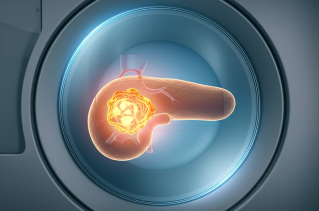
Pancreatic neuroendocrine neoplasms (PanNENs) are tumors arising from neuroendocrine cells in the pancreas. These tumors account for a small percentage of all pancreatic neoplasms, but their behavior and treatment strategies vary significantly. Accurate classification is crucial for determining the best course of action.

The World Health Organization (WHO) classification divides PanNENs into three grades: Grade 1 (G1), Grade 2 (G2), and Grade 3 (G3), also known as pancreatic neuroendocrine carcinoma (PanNEC). This grading is based on factors like mitotic count and Ki-67 proliferation index, which reflect how quickly the tumor cells are dividing. Higher grades indicate more aggressive tumors.

Traditional methods of detection include endoscopic ultrasound, computed tomography (CT), magnetic resonance imaging (MRI), and positron emission tomography (PET)/CT. This article will focus on how MRI can play a crucial role in distinguishing between PanNETs G1/G2 and the more aggressive PanNEC G3, guiding treatment decisions before surgical intervention.

MRI: A Non-Invasive Tool for Grading Pancreatic Tumors

MRI scan revealing a pancreatic tumor.

Magnetic resonance imaging (MRI) is a non-invasive imaging technique that provides detailed information about the characteristics of PanNENs. MRI can help assess tumor location, size, boundary, and internal composition, as well as detect any spread to nearby tissues or distant organs. By analyzing these features, radiologists can gain insights into the tumor's aggressiveness and guide treatment decisions.

A recent study published in OncoTargets and Therapy investigated the MRI features of PanNENs in 59 patients. The study compared the MRI characteristics of PanNETs G1/G2 with those of PanNEC G3, aiming to identify features that could differentiate between the two groups. Researchers carefully analyzed various MRI parameters, including:

  • Tumor location
  • Size
  • Boundary (well-defined vs. ill-defined)
  • Appearance (cystic vs. solid)
  • Enhancement patterns after contrast injection
  • Pancreatic duct dilatation
  • Presence of metastases
  • Signal intensity on different MRI sequences
  • Apparent diffusion coefficient (ADC) values
The study revealed several significant differences between PanNETs G1/G2 and PanNEC G3 on MRI. PanNEC G3 tumors were more likely to exhibit ill-defined boundaries, larger sizes, necrosis, lower enhancement, pancreatic duct dilatation, metastases, and high signal intensity on diffusion-weighted imaging (DWI). Additionally, PanNEC G3 tumors had significantly lower ADC values compared to PanNETs G1/G2 and normal pancreatic tissue.

The Future of Pancreatic Tumor Diagnosis

MRI is a valuable tool for differentiating between PanNEC G3 and PanNETs G1/G2, which can ultimately guide treatment decisions and improve patient outcomes. By identifying key MRI features and ADC value thresholds, radiologists can provide crucial information to surgeons and oncologists, leading to more personalized and effective treatment strategies.

About this Article -

This article was crafted using a human-AI hybrid and collaborative approach. AI assisted our team with initial drafting, research insights, identifying key questions, and image generation. Our human editors guided topic selection, defined the angle, structured the content, ensured factual accuracy and relevance, refined the tone, and conducted thorough editing to deliver helpful, high-quality information.See our About page for more information.

Everything You Need To Know

1

What are Pancreatic Neuroendocrine Neoplasms (PanNENs), and why is it important to classify them correctly?

Pancreatic Neuroendocrine Neoplasms (PanNENs) are tumors that originate from the neuroendocrine cells in the pancreas. Correct classification is crucial because PanNENs exhibit varying degrees of aggressiveness, and the appropriate treatment strategy depends on accurately determining whether a tumor is a well-differentiated PanNET Grade 1 (G1) or Grade 2 (G2), or a more aggressive Pancreatic Neuroendocrine Carcinoma (PanNEC) Grade 3 (G3). This classification guides treatment decisions, potentially impacting patient outcomes. Other characterizations, for example by stage, are also useful to guide treatment planning.

2

How does the World Health Organization (WHO) classify PanNENs, and what factors are considered in this classification?

The World Health Organization (WHO) classifies PanNENs into three grades: Grade 1 (G1), Grade 2 (G2), and Grade 3 (G3), with Grade 3 also referred to as Pancreatic Neuroendocrine Carcinoma (PanNEC). This grading is primarily based on the mitotic count and Ki-67 proliferation index, which are measures of how quickly the tumor cells are dividing. Higher grades, like G3, signify more aggressive tumors, necessitating more aggressive treatment approaches.

3

What role does magnetic resonance imaging (MRI) play in the diagnosis and treatment of PanNENs?

Magnetic resonance imaging (MRI) is a non-invasive imaging technique that helps in assessing Pancreatic Neuroendocrine Neoplasms (PanNENs). MRI provides detailed information about tumor location, size, boundary characteristics (well-defined versus ill-defined), internal composition (cystic versus solid), enhancement patterns after contrast injection, and the presence of metastases. Furthermore, MRI can detect pancreatic duct dilatation and assess signal intensity on different MRI sequences, including apparent diffusion coefficient (ADC) values. By analyzing these features, radiologists can distinguish between PanNETs G1/G2 and the more aggressive PanNEC G3, guiding treatment decisions before surgical intervention. However, MRI is not the only tool used; other methods like endoscopic ultrasound, computed tomography (CT), and positron emission tomography (PET)/CT also contribute to the overall diagnostic picture.

4

What are the key MRI features that differentiate PanNEC G3 tumors from PanNETs G1/G2?

On MRI, PanNEC G3 tumors tend to exhibit several distinct features compared to PanNETs G1/G2. These include ill-defined boundaries, larger sizes, the presence of necrosis, lower enhancement after contrast injection, pancreatic duct dilatation, metastases, and high signal intensity on diffusion-weighted imaging (DWI). Additionally, PanNEC G3 tumors typically demonstrate significantly lower apparent diffusion coefficient (ADC) values when compared to PanNETs G1/G2 and normal pancreatic tissue. These differences enable radiologists to distinguish between the tumor types.

5

What are the implications of using MRI to differentiate between PanNEC G3 and PanNETs G1/G2 for patient treatment and outcomes?

Using magnetic resonance imaging (MRI) to differentiate between PanNEC G3 and PanNETs G1/G2 allows for more personalized and effective treatment strategies. By identifying key MRI features and ADC value thresholds, radiologists can provide critical information to surgeons and oncologists, leading to better-informed treatment decisions. This can result in improved patient outcomes, as the treatment approach is tailored to the specific characteristics and aggressiveness of the tumor, rather than applying a one-size-fits-all method. Earlier detection and characterization using MRI, combined with other diagnostic tools, can also optimize surgical planning and potentially improve survival rates. The potential benefit is that patients with more aggressive tumors can be identified earlier and start potentially more aggressive treatment sooner.

Newsletter Subscribe

Subscribe to get the latest articles and insights directly in your inbox.