Decoding Neuron Development: How Fruit Flies Can Help Us Understand Our Brains
"Researchers are using the humble fruit fly to unlock the secrets of neuron development, offering new insights into how our brains are wired and what happens when things go wrong."
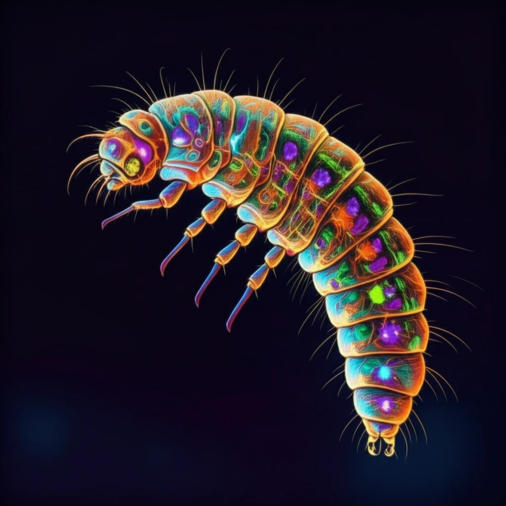
The development of a nervous system is an intricate process, requiring precise neuron positioning and identity, followed by specific dendritic development and axonal wiring. Recently, researchers have turned to the dendritic arborization (DA) sensory neurons of Drosophila larvae, commonly known as fruit flies, as a powerful genetic model. These neurons offer a unique opportunity to study the mechanisms that govern neuron differentiation, providing insights applicable to more complex organisms.
Drosophila's DA neurons are categorized into four main classes (I-IV), each with increasing dendrite complexity and distinct genetic controls. This classification allows scientists to investigate the molecular mechanisms behind dendritic morphology. The advantages of using fruit flies include the availability of advanced genetic tools, the two-dimensional spread of dendrite arbors beneath a transparent larval cuticle for easy visualization, and the diverse dendritic morphologies that facilitate comparative analysis. Additionally, the stereotypical shapes of different DA neuron arbors enable detailed morphometric statistical analyses.
The activity of DA neurons influences larval locomotion, with each class possessing distinct sensory modalities that trigger different behavioral responses. Axonal projections from these neurons extend into the larval central nervous system (VNC), forming topographic representations of sensory modality and body wall position. Studying these axonal projections can reveal mechanisms underlying topographic mapping and the wiring of circuits that modulate larval locomotion.
Genetic Mosaic Analysis: A Powerful Tool for Neuron Study

Genetic mosaic analysis, particularly using techniques like MARCM (Mosaic Analysis with a Repressible Cell Marker) and Flp-out, allows researchers to mark and analyze DA neurons in Drosophila larvae. These methods enable the creation of genetically distinct populations of cells within the same organism, facilitating the study of gene function in specific neurons.
- Reagent Preparation: Essential reagents include calcium-free HL3.1 saline to prevent muscle contraction during dissection and poly-L-lysine (PLL)-coated coverslips for mounting samples.
- Genetic Crosses: Specific genetic crosses are performed to generate MARCM or Flp-out clones. For example, generating MARCM clones involves crossing FRT2A flies with hsFLP; Gal4109(2)80, UAS-mCD8::GFP; tub-Gal80 FRT2A/SM5-TM6B flies. For Flp-out clones, a cross might involve ppk-Gal4 flies with yw, hsFLP;UAS-FRT-CD2,y-stop-FRT-mCD8::GFP flies.
- Embryo Collection and Heat Shock: Embryos are collected and subjected to heat shock treatments to induce the expression of specific genes. The duration and temperature of the heat shock are critical for controlling the frequency of clone generation.
- Clone Screening: After heat shock, embryos are cultured until they reach the wandering 3rd instar larval stage. Larvae with GFP-positive neurons/cells are identified using a fluorescence dissecting microscope.
- In Vivo Imaging: Live larvae are placed in a depression slide with glycerol and imaged using confocal microscopy to visualize the dendritic arbor of mCD8::GFP-labeled neurons.
- Larval Dissection: Dissection is performed rapidly (within 5 minutes per larva) to preserve dendrite morphology. The central nervous system (CNS) is kept intact when imaging DA neuron axon termini.
- Fixation and Blocking: Larval fillets are fixed in 4% PFA, washed, and blocked with normal donkey serum (NDS) to prepare them for antibody staining.
- Staining: Larval fillets are incubated with primary and secondary antibodies to label specific neuronal structures.
- Mounting: Larval fillets are mounted on slides for examination of dendrite arbors or axon termini. Special techniques ensure the samples are flat and well-preserved.
The Future of Neuron Research
The Drosophila larval DA neuron model provides a powerful system for investigating the mechanisms controlling neuron morphology and circuit formation. By combining genetic mosaic analysis with advanced imaging techniques, researchers can gain insights into the development of the nervous system and identify potential targets for therapeutic interventions. These findings contribute to our understanding of brain function and offer new avenues for treating neurological disorders.
