Chick embryo developing ex ovo in a sterile lab environment, symbolizing advancements in developmental biology.

Cracking the Egg: Revolutionizing Chick Embryo Research for Advances in Developmental Studies

"A streamlined, shell-less culturing technique opens new doors for observing and manipulating chick embryos, pushing the boundaries of developmental biology."


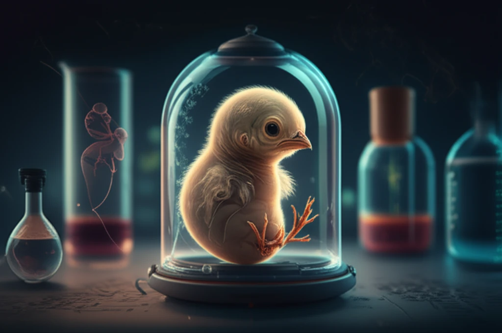
Research in anatomy, embryology, and developmental biology relies heavily on model organisms. Chick embryos are a popular choice due to their low cost and ease of maintenance. However, the opaque eggshell presents challenges for observation. To overcome this, researchers have developed techniques such as windowing and ex-ovo culturing to view the embryo as it develops.

Both windowing and ex-ovo techniques have their own advantages and limitations. The windowing method involves creating a hole in the eggshell, while ex-ovo culturing removes the embryo from the shell entirely. Each method presents unique challenges, driving the need for optimized approaches.

This article introduces an improved and simplified ex-ovo culturing technique for chick embryos. This method enables observation of embryonic development from stage HH 19 into later stages (HH 40), allowing researchers to study organ development. This technique is easily adaptable for both undergraduate classes and advanced research laboratories.

Ex-Ovo Culturing: Solving Key Challenges in Embryo Development Research

Chick embryo developing ex ovo in a sterile lab environment, symbolizing advancements in developmental biology.

Traditional ex-ovo techniques often involve using a Styrofoam cup or glass bowl lined with plastic wrap. While effective, this setup can be technically challenging. The plastic wrap is difficult to manage, often slipping or tearing, posing a risk to the embryo. Additionally, the setup is not very stable and can be easily knocked over, and the height of the cups can make it difficult to place the embryo under a stereomicroscope.

In contrast, the windowing method creates a hole in the eggshell to view the embryo. While this allows for easy maintenance and observation, large windows are difficult to seal, leading to sterility and survivability problems. The process of using molten wax as a sealant can also be inconvenient and messy.

Here’s how this improved ex ovo culturing technique can benefit researchers and students:
  • Eliminates the need for high-tech equipment.
  • Simplifies handling under a stereomicroscope.
  • Provides ample support for microscopic manipulations.
  • Enables complete viewing of the embryo into later stages of development (up to HH 40-41).
The benefits of this ex ovo technique are substantial, particularly for observing developmental processes that occur later in development, such as ossification, feather development, and advanced limb and eye development. This method allows for clear visualization of the entire embryo, extraembryonic membranes, and vasculature. Researchers can also perform advanced procedures like embryonic manipulations, gaining insights into the effects of these manipulations on later-stage embryos.

The Future of Chick Embryo Research

In conclusion, the optimized ex ovo method provides an ideal solution for viewing and manipulating chick embryos, offering unrestricted access and extending the observable stages of development. By addressing the challenges associated with traditional methods, this technique enhances research capabilities and educational opportunities in developmental biology. Researchers and students will appreciate the simplicity, stability, and accessibility it provides, paving the way for new discoveries and a deeper understanding of embryonic development.

About this Article -

This article was crafted using a human-AI hybrid and collaborative approach. AI assisted our team with initial drafting, research insights, identifying key questions, and image generation. Our human editors guided topic selection, defined the angle, structured the content, ensured factual accuracy and relevance, refined the tone, and conducted thorough editing to deliver helpful, high-quality information.See our About page for more information.

Everything You Need To Know

1

What is ex-ovo culturing, and why is it important for chick embryo research?

Ex-ovo culturing is a method used in developmental biology research where a chick embryo is removed from its eggshell and grown in an artificial environment. This is significant because the opaque eggshell normally obscures the view of the developing embryo. By using ex-ovo culturing, researchers can directly observe and manipulate the embryo during its development. This allows for detailed study of organ development, vascularization, and the effects of various experimental manipulations. Without this, studying these processes would be much more difficult, limiting advancements in developmental biology.

2

What are Hamburger-Hamilton (HH) stages, and why is it significant to reach HH 40-41 in chick embryo research?

The Hamburger-Hamilton (HH) stages are a standardized system used to classify the developmental progress of chick embryos. Reaching stage HH 40-41 is significant because it represents a relatively late stage in embryonic development where many critical processes, such as organogenesis, ossification, and the development of feathers and limbs, are well underway. Being able to observe chick embryos up to HH stage 40-41 provides valuable insights into the later stages of development, which are crucial for understanding congenital abnormalities and developmental processes.

3

How does the windowing method compare to ex-ovo culturing in chick embryo research?

The windowing method involves creating a small opening in the eggshell to observe the chick embryo. This contrasts with ex-ovo culturing, where the embryo is completely removed from the shell. Windowing allows for observation while maintaining a more natural environment for the embryo, but it can be challenging to maintain sterility and seal the window properly. Ex-ovo culturing provides better access for manipulation and clearer visualization, but it requires a more controlled environment to ensure the embryo's survival. Both methods have their advantages and are chosen depending on the specific needs of the experiment.

4

Why are stereomicroscopes important in chick embryo research, and how does the culturing technique help?

Stereomicroscopes are essential tools in chick embryo research because they allow researchers to view the embryo in three dimensions at relatively low magnification. This is particularly important for performing delicate manipulations, observing surface details, and documenting developmental progress. An optimized culturing technique that simplifies handling under a stereomicroscope enables more precise and efficient research, leading to better data and more reliable results. Improved techniques also enable better access and manipulation of the embryo when viewing through the lens of the microscope.

5

What are the challenges of traditional ex-ovo culturing methods, and how do improved techniques address these issues?

Traditional ex-ovo culturing methods often involve using simple setups like Styrofoam cups and plastic wrap. While these methods can be effective, they also come with challenges such as instability, difficulty in handling the plastic wrap, and limited visibility. Improved ex-ovo culturing techniques aim to address these issues by providing a more stable, accessible, and easily manageable environment for the chick embryo. This not only improves the success rate of the culturing but also makes the technique more accessible for both research and educational purposes. This can significantly enhance research capabilities and improve the learning experience.

Newsletter Subscribe

Subscribe to get the latest articles and insights directly in your inbox.