
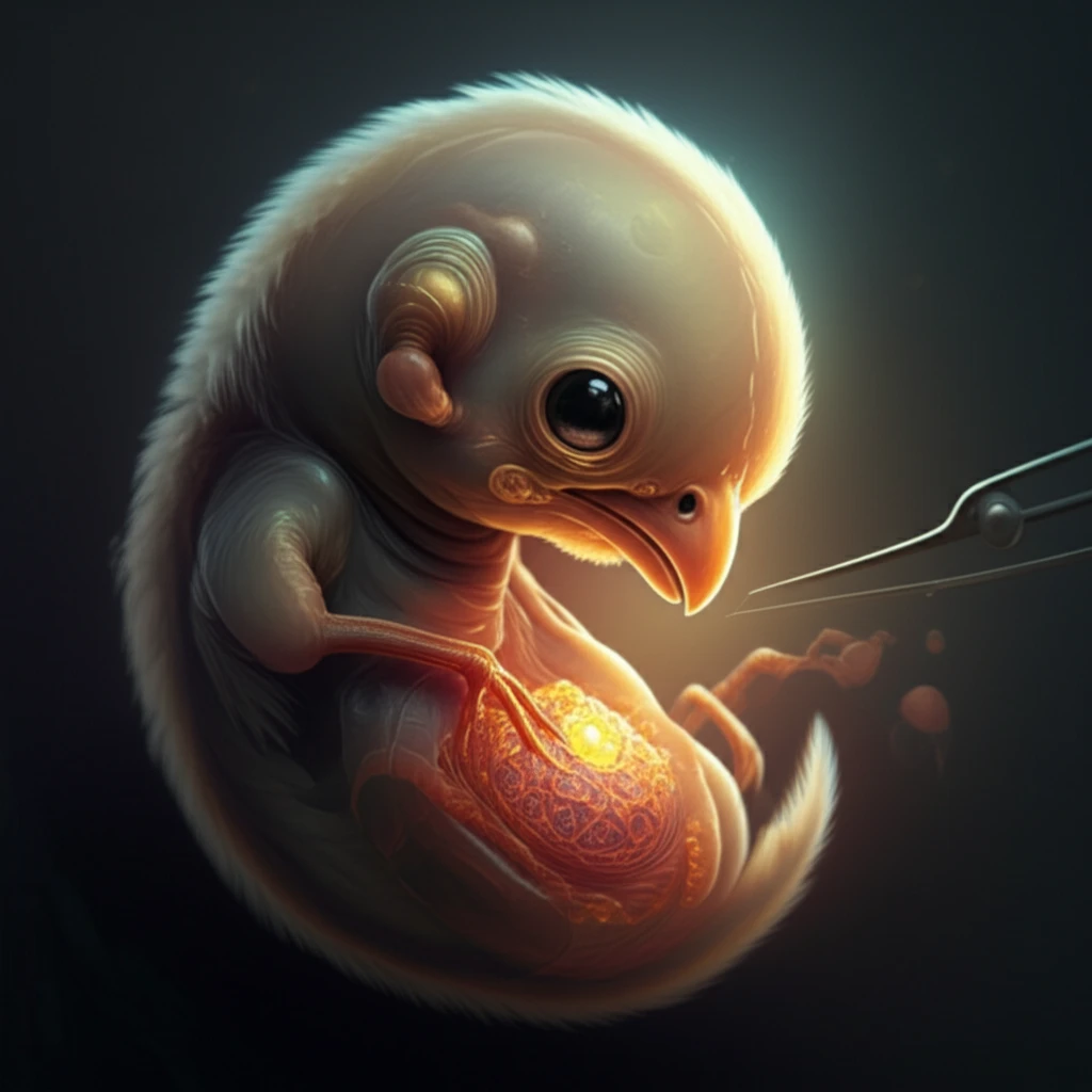
Unlocking Facial Development: How Avian Embryos Reveal Secrets of the Face

"By transplanting tissues between quail and chick embryos, scientists gain insights into the signaling processes that shape our faces."
The study of embryonic development has long been aided by the accessibility of avian embryos. These embryos allow scientists to perform experiments that reveal how cells differentiate and how tissues interact to form complex structures, especially in vertebrates.
One powerful technique involves creating quail-chick or mouse-chick chimeras. This is achieved by transplanting ectodermal tissue (the outer layer of cells) from a quail or mouse embryo to a chick embryo. Since quail cells possess a unique nucleolar marker and mouse cells have a specific repetitive element, researchers can easily distinguish donor from host tissue.
This method is particularly useful for studying facial development. By transplanting ectoderm from the upper jaw region of a quail or mouse embryo onto a chick embryo, scientists can investigate the signaling properties of this tissue and how it influences the development of the face.
The Scope of Craniofacial Development Research
Craniofacial anomalies represent a significant category of birth defects affecting facial and skull formation. While precise global prevalence figures vary by condition, these developmental disorders collectively impact a substantial portion of live births worldwide. Understanding the fundamental mechanisms of facial development remains a priority for both basic science and clinical research.
Traditional Models in Craniofacial Research
Animal models have long served as the foundation for studying craniofacial development, with avian and mammalian embryos commonly employed. These models offer experimental accessibility and developmental similarities to humans, though extrapolating findings across species requires careful validation. Limitations include differences in gestational environments and the inherent complexity of translating basic developmental biology to clinical applications.
Evolution of Avian Embryo Research
The chicken embryo emerged as a premier model organism for embryological research over a century ago, valued for its accessibility and rapid development. Landmark studies established avian embryos as powerful tools for understanding vertebrate craniofacial morphogenesis. These foundational contributions continue to shape contemporary research approaches in developmental biology.
How Tissue Transplants Illuminate Facial Development
The researchers carefully dissect the frontonasal process (the precursor to the upper jaw) from a quail or mouse embryo. This tissue is then treated with dispase, an enzyme that separates the ectoderm from the underlying mesenchyme and neuroectoderm.
- The donor ectoderm is then transplanted into this space and secured with glass pins.
- The embryo is allowed to develop, and the researchers analyze the distribution of donor and host tissues using specific markers.
- This allows them to determine how the transplanted ectoderm influences the surrounding tissues and the overall development of the face.
Advances in Craniofacial Developmental Studies
Recent research has increasingly focused on understanding the precise mechanisms governing craniofacial morphogenesis during embryonic development. Contemporary investigations employ sophisticated imaging and molecular techniques to dissect developmental pathways. These studies contribute to a growing body of knowledge regarding the genetic and environmental factors influencing facial formation.
Challenges in Avian-Human Translational Research
Translating findings from avian models to human craniofacial development faces inherent biological and methodological challenges. Critics note that while avian embryos share fundamental developmental processes with mammals, significant species-specific differences exist. The field continues to grapple with establishing the precise boundaries of cross-species applicability.
Integrating Evidence Across Model Systems
Comparative studies examining craniofacial development across multiple species have yielded insights into both conserved and divergent mechanisms. The integration of avian, rodent, and other model system data provides a more comprehensive understanding of developmental principles. This multi-model approach helps identify which findings represent universal biological processes versus species-specific adaptations.
Unraveling the Secrets of Facial Formation
This transplantation method has revealed that the ectoderm plays a crucial role in regulating dorsoventral polarity and proximodistal extension of the upper jaw. The fact that similar results are obtained with both quail and mouse ectoderm suggests that these signaling mechanisms are highly conserved across species.
Key Findings on Eye-Face Developmental Connections
Research on chicken embryos has revealed important insights about the relationship between developing eyes and craniofacial structures. A 2020 study published in Mechanisms of Development specifically investigated whether embryonic eyes exert influence over craniofacial prominence development and morphology at early stages. Additionally, work by Brugmann et al. (2006) demonstrated that more distantly related avian species show species-specific craniofacial morphologies earlier in embryogenesis, while closely related species maintain facial similarity longer during development. These findings underscore the value of avian models in understanding the timing and mechanisms of facial morphological divergence.
Emerging Directions in Avian Craniofacial Research
Future investigations will likely leverage advancing technologies to probe craniofacial development with unprecedented resolution. Integrating genomic, imaging, and computational approaches promises to deepen mechanistic understanding. Continued refinement of avian models may reveal novel therapeutic targets for craniofacial disorders.
Developmental Trajectories and Evolutionary Constraints
The hourglass model of development suggests that while early developmental events may be highly evolvable, a conserved phylotypic stage exists across vertebrates. Research on craniofacial developmental trajectories demonstrates how morphological divergence among related species involves changes to developmental processes, with significant theoretical interest in when such variation arises. A 2025 study in Proceedings of the Royal Society B further emphasizes that avian cranial evolution is influenced by shape interactions among cranial elements, with the brain playing a key role in development of surrounding tissues while other cranial elements also moderate morphological evolution. These findings highlight the complex interplay between developmental constraints and evolutionary divergence in shaping craniofacial diversity.
Translating Avian Research to Human Health
Understanding craniofacial development in avian models holds potential implications for addressing human birth defects and developmental disorders. While direct clinical applications remain in development, the fundamental biological insights gained from avian research contribute to the broader knowledge base supporting eventual therapeutic advances. Continued investment in basic developmental biology research remains essential for long-term medical progress.
Furthermore, this technique can be used to identify multiple signaling centers within the ectoderm that contribute to facial morphogenesis. By combining the strengths of mouse genetics with the avian-chimera system, researchers can identify the specific molecules that mediate epithelial-mesenchymal interactions during development.
Ultimately, a deeper understanding of these interactions will pave the way for new approaches to prevent and treat craniofacial malformations, improving the lives of individuals affected by these conditions.