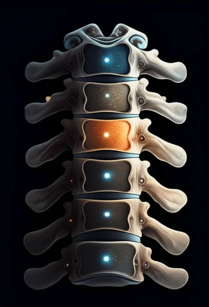
Cracking the C2 Code: How a Tiny Foramen Can Improve Spinal Surgery
"Unlock the secrets of the C2 axis with a surprising surgical landmark – nutrient foramina. New research reveals how these tiny openings can enhance pedicle screw placement and patient outcomes."
Surgeons face a unique challenge when placing pedicle screws into the second cervical vertebra (C2). The C2 vertebra's complex anatomy requires precision to avoid complications. While surgeons use various landmarks and measurements, the lack of universal consensus in existing literature often leads to variability in technique and outcomes.
Innovative anatomical research is exploring a new surgical landmark: the nutrient foramina. These tiny holes in the posterior aspect of C2 allow blood vessels to enter the bone. A recent study investigates the potential of nutrient foramina as a consistent and reliable guide for C2 pedicle screw placement.
This article breaks down the study's findings, explaining how surgeons can use nutrient foramina to improve the accuracy and safety of C2 pedicle screw placement. We'll explore the methods used, the key measurements revealed, and the clinical implications of this anatomical approach. This innovative landmark will help you revolutionize spinal surgery.
Nutrient Foramina: A Surgeon's New Best Friend?

Researchers conducted a detailed anatomical study using 19 dry bone C2 specimens (38 sides) and one cadaveric specimen. The study carefully documented the presence, size, location, and distance from the midline of the nutrient foramina, found where the isthmus and lamina meet. Dissection of the cadaveric specimen helped reveal the source of arteries entering these foramina.
- The number of foramina ranged from 0 to 5, with an average of 1.84 per side.
- About 7.9% of sides did not have any foramina at all.
- The average diameter of the foramina was 0.57 mm.
- The foramina were most commonly found in position 2 (66.4% of sides), followed by position 3 (24.1%) and position 1 (9.5%).
- The average horizontal distance from the midline of the spinous process to the foramina was 25.17 mm.
- In the cadaveric specimen, the arteries entering the C2 nutrient foramina originated from distal branches of the deep cervical artery.
Better Surgery Through Better Landmarks
This anatomical study provides valuable insights into using nutrient foramina as a landmark for C2 pedicle screw placement. By understanding the average location, size, and potential absence of these foramina, surgeons can improve the accuracy and safety of their procedures.
While technological advancements like navigation-guided placement of pedicle screws exist, anatomical landmarks remain crucial, especially when advanced technology isn't available. This study supports combining traditional anatomical knowledge with modern techniques for optimal surgical outcomes.
The research underscores the importance of detailed anatomical knowledge in spinal surgery. As the study highlights, variations exist, making a thorough understanding of individual anatomy crucial for success. This knowledge empowers surgeons to refine their techniques and personalize their approach, ultimately improving patient care.
