Chest Tube Training on a Budget: How a Swine Model Can Save Lives
"Discover how a low-cost, readily available swine model is revolutionizing chest tube insertion training, making life-saving skills accessible to medical professionals everywhere."
In the high-stakes world of medicine, proficiency in life-saving procedures is paramount. Chest tube insertion, a critical intervention for conditions like collapsed lungs or traumatic chest injuries, demands precision and expertise. Traditionally, medical training has relied on expensive simulation models or, less desirably, direct patient practice. But what if there was a more accessible, cost-effective way to master this essential skill?
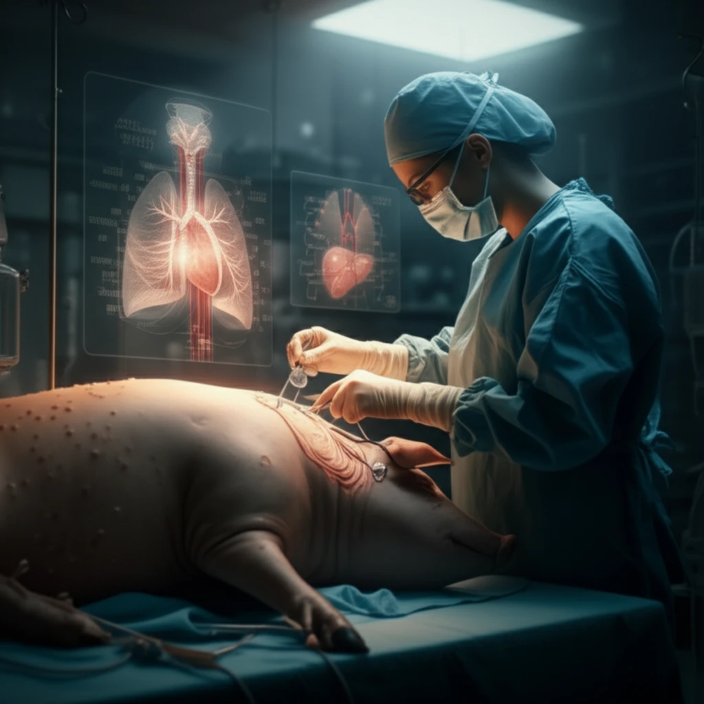
Enter the innovative swine model. A recent study out of Paraná, Brazil, highlights the effectiveness of using readily available porcine (pig) tissue to simulate the human chest cavity. This low-cost, low-technology approach offers a compelling alternative for medical students and residents to hone their skills in a realistic and controlled environment. This is especially relevant as medical educators strive to balance effective training with ethical and financial considerations.
This article delves into the details of this groundbreaking teaching project, exploring how the swine model was developed, how it was used in training, and why it's gaining traction as a valuable tool in medical education. We'll uncover the benefits, address the limitations, and discuss the potential for this accessible method to improve patient outcomes worldwide.
Why Swine? The Benefits of a Low-Cost Training Model
The appeal of the swine model lies in its simplicity and affordability. Unlike sophisticated mannequins that can cost thousands of dollars, porcine rib cages can be sourced from local food markets at a fraction of the price. This makes high-quality medical training more accessible to institutions with limited budgets, particularly in developing countries.
- Realistic Simulation: The texture and structure of porcine tissue closely mimic that of the human chest wall, providing a realistic tactile experience for trainees.
- Hands-On Practice: Medical students and residents can perform the entire chest tube insertion procedure, from incision to tube placement, under the guidance of experienced instructors.
- Safe Learning Environment: The controlled setting of the training lab eliminates the risk of patient harm during the initial learning stages.
- Ethical Considerations: Using animal byproducts reduces reliance on live animal models or cadavers, addressing ethical concerns related to animal welfare and human remains.
- Versatile Training: The model can be adapted to simulate various clinical scenarios, such as traumatic injuries or fluid accumulation in the chest cavity.
The Future of Medical Training: Accessible, Affordable, and Effective
The success of the swine model in chest tube insertion training highlights the potential for innovative, low-cost solutions to transform medical education. By embracing readily available resources and prioritizing hands-on learning, medical institutions can empower the next generation of healthcare professionals with the skills and confidence they need to save lives. As the study authors concluded, this model has potential use as a teaching tool in medical education, especially in resource-limited settings.