Illustration of activated neutrophils releasing vacuoles in a blood vessel, with hematology analyzer data in the background.

Can Your Blood Test Reveal Hidden Infections? A New Way to Spot Neutrophil Activation

"Scientists have discovered a novel method to detect activated neutrophils using standard hematology analyzers, potentially offering earlier insights into bacterial infections and immune responses."


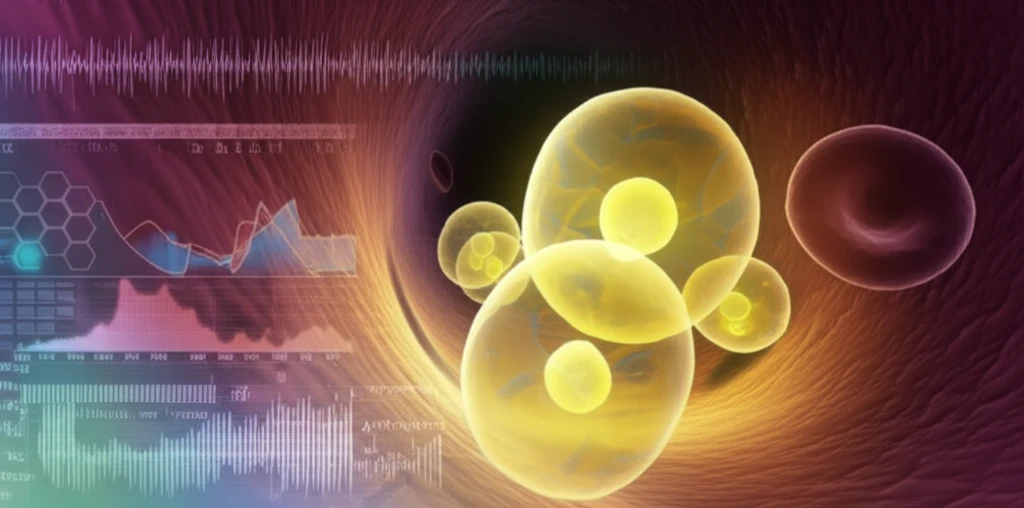
Our bodies have a sophisticated defense system, and at the forefront of that system are neutrophils, a type of white blood cell. These cells are essential for fighting off infections, primarily by engulfing bacteria (phagocytosis) and releasing reactive oxygen species (ROS) to kill pathogens. When an infection occurs, the number of neutrophils increases, and they're called to the site of infection. Understanding how neutrophils function and respond is crucial for managing various health conditions.

Traditionally, identifying activated neutrophils, which are neutrophils that are actively fighting infection, has been complex, often involving specialized imaging techniques. However, a recent study sheds light on a simpler approach. Researchers have found that by using standard hematology analyzers—machines commonly used for routine blood tests—it’s possible to detect activated neutrophils based on their unique characteristics.

This article explores this innovative method, explaining how changes in neutrophil structure, specifically the formation of vacuoles, can be detected using ordinary blood testing equipment. This approach offers the potential for quicker and more accessible diagnosis of bacterial infections and a better understanding of how our immune system responds to threats.

Vacuoles: The Key to Unlocking Neutrophil Activity

Illustration of activated neutrophils releasing vacuoles in a blood vessel, with hematology analyzer data in the background.

The key to this new detection method lies in the observation that activated neutrophils form vacuoles—small, bubble-like structures—within their cytoplasm. These vacuoles are created as part of the process where neutrophils engulf and destroy pathogens. The presence of these vacuoles changes the way light scatters when analyzed by a hematology analyzer, particularly increasing side-scattering (SSC).

To investigate this, researchers activated neutrophils with substances like phorbol myristate acetate (PMA) and formylmethionylleucylphenylalanine (fMLP), which mimic bacterial signals. They then used a Sysmex XE-2100 automated hematology analyzer to observe the changes in these cells. What they found was significant:

  • A distinct cluster of cells appeared in the eosinophil area of the analyzer's scattergram, indicating a population with higher side-scattering (SSC).
  • These cells were confirmed to be CD16b- and APF-positive, markers of activated neutrophils producing ROS.
  • Microscopic analysis (CLSM and EM) revealed the presence of vacuoles within these activated neutrophils.
This means that the increased side-scattering observed by the hematology analyzer directly correlates with the presence of vacuoles in activated neutrophils. This correlation provides a new way to quickly identify these infection-fighting cells using readily available technology.

The Future of Infection Detection

This research opens up exciting possibilities for diagnosing and monitoring bacterial infections. By using standard hematology analyzers to detect changes in neutrophil structure, doctors may be able to identify infections earlier and more efficiently.

Furthermore, this method could improve our understanding of various immune responses. Since neutrophil activation plays a key role in many inflammatory conditions, this technique could be used to monitor disease activity and response to treatment.

While further studies are needed to fully validate this approach, the initial findings are promising. This innovative use of existing technology has the potential to transform how we detect and manage infections, ultimately leading to better patient outcomes.

About this Article -

This article was crafted using a human-AI hybrid and collaborative approach. AI assisted our team with initial drafting, research insights, identifying key questions, and image generation. Our human editors guided topic selection, defined the angle, structured the content, ensured factual accuracy and relevance, refined the tone, and conducted thorough editing to deliver helpful, high-quality information.See our About page for more information.

This article is based on research published under:

DOI-LINK: 10.1016/j.jim.2018.10.004, Alternate LINK

Title: Detection Of Activated Neutrophils By Reactive Oxygen Species Production Using A Hematology Analyzer

Subject: Immunology

Journal: Journal of Immunological Methods

Publisher: Elsevier BV

Authors: Mari Kono, Katsuyasu Saigo, Shiori Matsuhiroya, Takayuki Takahashi, Makoto Hashimoto, Ayako Obuchi, Shion Imoto, Takashi Nishiyama, Seiji Kawano

Published: 2018-12-01

Everything You Need To Know

1

What are neutrophils, and why are they important?

Neutrophils are a type of white blood cell, crucial in the body's defense system. Their primary function is to fight off infections, mainly by engulfing bacteria through phagocytosis and releasing reactive oxygen species (ROS) to kill pathogens. Neutrophils are essential for managing various health conditions because they are at the forefront of our immune response, and understanding their function is critical for diagnosing and treating infections.

2

What are activated neutrophils?

Activated neutrophils are neutrophils that are actively engaged in fighting an infection. They exhibit specific characteristics, such as forming vacuoles within their cytoplasm. This activation process is essential for initiating an immune response to combat bacterial infections. The presence of these vacuoles and other changes within activated neutrophils can be detected using standard hematology analyzers.

3

How can activated neutrophils be detected using a hematology analyzer?

Researchers have discovered a new method to detect activated neutrophils using standard hematology analyzers. These machines, commonly used for routine blood tests, can identify activated neutrophils by detecting changes in their structure, specifically the formation of vacuoles. The analyzers measure how light scatters when it interacts with the cells. The presence of vacuoles in activated neutrophils alters the light scattering, which the analyzer then detects as increased side-scattering (SSC). This method offers a quicker and more accessible approach for diagnosing bacterial infections.

4

What are vacuoles, and what role do they play in this new detection method?

Vacuoles are small, bubble-like structures that form within the cytoplasm of activated neutrophils. They appear during the process where neutrophils engulf and destroy pathogens. The presence of these vacuoles significantly changes how light scatters when analyzed by a hematology analyzer, increasing side-scattering (SSC). The formation of vacuoles is a key indicator of neutrophil activation and their role in fighting infections. Detecting vacuoles is how the new method is able to quickly identify these infection-fighting cells.

5

What is the importance of this new method for infection detection?

The significance of this research lies in its potential for earlier and more efficient diagnosis of bacterial infections. By utilizing standard hematology analyzers, doctors may identify infections sooner, allowing for quicker and more effective treatment. This can lead to improved patient outcomes and a better understanding of the immune system's response to threats. The ability to quickly detect activated neutrophils also enables monitoring of treatment efficacy and the progression of infections, offering a new way to understand and manage infections.

Newsletter Subscribe

Subscribe to get the latest articles and insights directly in your inbox.