AI-powered surgical tool examining bowel

Can AI Spot Bowel Problems Better Than Surgeons? The Future of Gut Checkups

"New research explores how computer-assisted analysis of fluorescent signals can distinguish between arterial and venous bowel ischemia, paving the way for more precise surgical interventions."


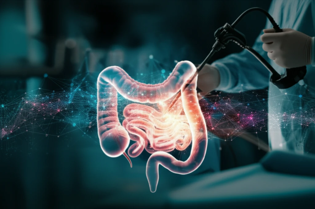
Imagine a world where surgeons have a high-tech assistant that can 'see' beneath the surface of tissues, instantly identifying problems that might otherwise be missed. That world is closer than you think, thanks to the innovative field of fluorescence angiography (FA). FA allows doctors to visualize blood flow in real time by injecting a fluorescent dye and using special cameras to capture the signal. This technique is increasingly used in surgeries to assess tissue viability, especially in the gut, where blood supply is critical for healing.

Bowel ischemia, a condition where the bowel doesn't receive enough blood, can lead to serious complications after surgery. The challenge? Distinguishing between arterial (inflow) and venous (outflow) problems. Current FA techniques often rely on a surgeon's subjective interpretation of the fluorescent signal, which can be influenced by factors like dye diffusion and camera angle. This is where the power of artificial intelligence (AI) comes in.

New research investigates how computer-assisted analysis of fluorescence signals can objectively differentiate between arterial and venous bowel ischemia. By using machine learning algorithms to analyze the dynamics of the fluorescent signal, this approach aims to provide a more accurate and reliable assessment of bowel perfusion, potentially leading to better surgical decisions and improved patient outcomes. This article will delve into this groundbreaking research, explaining how it works and what it could mean for the future of gut checkups.

How Does This AI-Powered 'Gut Check' Work?

AI-powered surgical tool examining bowel

The study, published in Surgical Endoscopy, involved creating different types of bowel ischemia in pigs: arterial (blocking the artery), venous (blocking the vein), and mixed (blocking both). Researchers then injected a fluorescent dye and used a special laparoscope (a camera inserted through a small incision) to capture the fluorescent signal. The key innovation was the use of a software called ER-PERFUSION, which analyzes the signal's intensity and speed to create a color-coded 'virtual perfusion cartography'.

This cartography provides a detailed picture of blood flow dynamics, highlighting areas of concern. But the analysis didn't stop there. The researchers also measured capillary lactate levels (a marker of tissue damage) in different regions of the bowel and fed all this data into machine learning algorithms. These algorithms were trained to recognize patterns associated with each type of ischemia.

Here's a breakdown of the key steps:
  • Creating Ischemia Models: Arterial, venous, and mixed ischemia were induced in pigs.
  • Fluorescence Imaging: A dye was injected, and a laparoscope captured the fluorescent signal.
  • Software Analysis: ER-PERFUSION software created a virtual perfusion cartography.
  • Data Collection: Capillary lactate levels were measured.
  • Machine Learning: Algorithms were trained to identify patterns of ischemia.
So, what did they find? The AI algorithms were able to distinguish between arterial and venous ischemia with impressive accuracy (up to 85% in some cases). The analysis of the fluorescent signal revealed distinct patterns for each type of ischemia. For example, venous ischemia showed a brighter initial signal and a faster time-to-peak compared to arterial ischemia. These findings suggest that AI-powered image analysis can provide a more objective and nuanced assessment of bowel perfusion than traditional methods.

The Future of Gut Checkups: What Does This Mean for You?

This research is an exciting step towards more precise and personalized surgical care. By providing surgeons with a high-tech 'gut check' tool, AI-powered image analysis could help reduce complications, improve patient outcomes, and even minimize the extent of bowel resections. Imagine a future where surgeons can confidently identify and address bowel perfusion problems in real-time, leading to faster recovery and a better quality of life for patients.

While this study is still in its early stages, the potential applications are vast. The technology could be used in a variety of surgical settings, including colorectal surgery, esophagectomy, and reconstructive surgery. It could also be adapted for use in other organs and tissues where blood supply is critical.

Of course, more research is needed to validate these findings in larger clinical trials. However, this study provides a compelling glimpse into the future of surgical care, where AI and human expertise work together to achieve the best possible outcomes for patients. As technology continues to advance, we can expect to see even more innovative tools emerge that will transform the way we diagnose and treat diseases of the gut.

About this Article -

This article was crafted using a human-AI hybrid and collaborative approach. AI assisted our team with initial drafting, research insights, identifying key questions, and image generation. Our human editors guided topic selection, defined the angle, structured the content, ensured factual accuracy and relevance, refined the tone, and conducted thorough editing to deliver helpful, high-quality information.See our About page for more information.

This article is based on research published under:

DOI-LINK: 10.1007/s00464-018-6512-6, Alternate LINK

Title: Discrimination Between Arterial And Venous Bowel Ischemia By Computer-Assisted Analysis Of The Fluorescent Signal

Subject: Surgery

Journal: Surgical Endoscopy

Publisher: Springer Science and Business Media LLC

Authors: Giuseppe Quero, Alfonso Lapergola, Manuel Barberio, Barbara Seeliger, Paola Saccomandi, Ludovica Guerriero, Didier Mutter, Alend Saadi, Marc Worreth, Jacques Marescaux, Vincent Agnus, Michele Diana

Published: 2018-10-16

Everything You Need To Know

1

How is Artificial Intelligence (AI) used in this new medical approach?

The application of AI in the medical field focuses on enhancing the diagnostic capabilities of surgeons in detecting and treating bowel ischemia. This involves using machine learning algorithms to analyze the data acquired through Fluorescence Angiography (FA) to differentiate between arterial and venous bowel ischemia. This technology is important because the ability to accurately distinguish between the types of ischemia allows for more targeted and effective surgical interventions, leading to improved patient outcomes. The use of AI in this context improves upon traditional methods by providing objective data analysis, reducing reliance on subjective interpretations.

2

What is Fluorescence Angiography (FA) and why is it important?

Fluorescence Angiography (FA) is a technique used to visualize blood flow in real time. A fluorescent dye is injected, and a special camera captures the signal. In the context of this topic, FA is crucial because it allows surgeons to assess the viability of tissues, especially in the gut, where blood supply is critical. Without FA, surgeons might have a harder time identifying areas of compromised blood flow, which could lead to complications. The implications of using FA are significant as it helps in making informed decisions during surgery, potentially preventing post-operative issues and improving recovery times.

3

What is bowel ischemia, and why is it important to distinguish between arterial and venous issues?

Bowel ischemia is a condition where the bowel doesn't receive enough blood, which can lead to serious complications. In the context of this topic, both arterial (inflow) and venous (outflow) problems are examined. Distinguishing between these types is vital because it determines how a surgeon will address the issue. Accurate identification leads to more effective treatments and minimizes the risk of complications. The implications of this are substantial, as misdiagnosis can lead to ineffective treatment and potentially worsen the patient's condition, emphasizing the need for precise diagnostic tools like those powered by AI.

4

What is ER-PERFUSION, and how does it contribute to this medical advancement?

ER-PERFUSION is a software used to analyze the fluorescent signal captured during Fluorescence Angiography (FA). The software creates a color-coded 'virtual perfusion cartography', providing a detailed picture of blood flow dynamics. The analysis also measured capillary lactate levels, a marker of tissue damage in different regions of the bowel, and all this data was fed into machine learning algorithms. These algorithms were trained to recognize patterns associated with each type of ischemia. ER-PERFUSION is crucial because it provides objective data analysis, assisting surgeons in making accurate assessments of bowel perfusion. The implications of using ER-PERFUSION are considerable, as it allows for a more comprehensive and objective evaluation of the condition compared to subjective interpretations.

5

How do machine learning algorithms improve the assessment of bowel perfusion?

Machine learning algorithms are used to analyze the data acquired during Fluorescence Angiography (FA) to differentiate between arterial and venous bowel ischemia. The AI algorithms analyze patterns in the fluorescent signal to distinguish between arterial and venous ischemia. This approach enhances the accuracy and reliability of assessing bowel perfusion. The implications of using machine learning are significant. It provides surgeons with a high-tech 'gut check' tool, potentially reducing complications, improving patient outcomes, and allowing for more precise surgical interventions, reducing the extent of bowel resections. This leads to faster recovery and a better quality of life for patients.

Newsletter Subscribe

Subscribe to get the latest articles and insights directly in your inbox.