
Breast Cancer Cell Lines: The Oxygen Connection You Need to Know

"Discover how growing breast cancer cells in more natural oxygen levels (5%) can change how we understand and treat this disease."
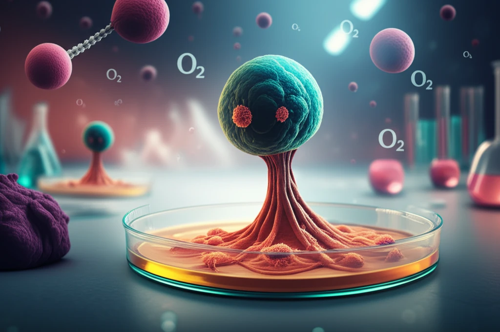
For years, most breast cancer cells studied in labs have been grown in air that's about 21% oxygen. This is much higher than the oxygen levels in our bodies, which usually average around 5%. This difference in oxygen can actually change how the cells behave, potentially skewing research results.
A new study is diving into this issue by growing breast cancer cells in a more realistic, low-oxygen (5%) setting. The goal is to see if these cells act differently and whether this could help us develop better ways to treat breast cancer.
These findings shed light on the importance of replicating true body conditions when studying cancer cells.
The Scale of Breast Cancer Cell Line Research
Breast cancer cell line research encompasses datasets of significant scale, with gene expression profiling studies analyzing hundreds of genes across dozens of cell lines—such as Affymetrix HG-U133A microarray studies of 39 breast cancer cell lines detailing 978 genes. Morphometric analyses have cataloged thousands of fluorescent images from panels of up to 41 breast cancer cell lines, enabling systematic subtyping. Murine models have also contributed, with hormone-sensitive and hormone-insensitive cell lines like MXT+ and MXT- established from the MXT-M-3,2 MC tumor model. These diverse datasets underscore the breadth of cellular models underpinning breast cancer research.
Cell Line Models and Their fidelity to Tumors
A central challenge in breast cancer research is determining how faithfully cell lines recapitulate the features of primary tumors, including epigenetic profiles such as DNA methylation. Researchers have evaluated methylation patterns across panels of breast cancer cell lines to assess their relevance as disease models. Beyond traditional monolayer cultures, researchers have developed miniature organ systems (organoids) to better predict how individual tumors respond to treatment. Intratumor heterogeneity—driven by the tumor microenvironment as well as genetic, epigenetic, and transcriptomic traits—further complicates the translation of cell line findings to patients. Understanding cell death mechanisms such as ferroptosis, an iron-dependent form of nonapoptotic cell death, adds another layer of complexity to therapeutic targeting.
Establishing Breast Cancer Cell Lines Across Ethnicities
The derivation of breast cancer cell lines from diverse sources represents a key milestone in cancer research. Cell lines such as IBH-4, IBH-6, and IBH-7 were established as novel hormone-responsive models, with some maintaining characteristics of the original tumor including drug resistance properties. Historically, most commonly used breast cancer cell lines originated from Caucasian or African American patients, with cell lines derived from other ethnic groups being comparatively rare. The recognition that BRCA1 and BRCA2 germline mutations, which are involved in DNA repair by homologous recombination and genomic stability, account for a large proportion of inherited breast cancer has also shaped which cell line models are developed. Established cell lines have been classified as basal- or luminal-like and as primary or metastatic based on their tumor subtype and origin.
The 5% Oxygen Advantage: What the Study Revealed
Scientists created four new breast cancer cell lines and grew them in 5% oxygen. They then compared these cells to standard cell lines grown in 21% oxygen, looking at various factors like:
- Three of the new cell lines (NZBR1, NZBR2, NZBR4) were triple-negative, meaning they didn't have estrogen receptors (ER-), progesterone receptors (PR-), or HER2. NZBR1 also had high levels of EGFR, a protein that can fuel cancer growth.
- One cell line (NZBR3) was HER2-positive and ER-positive, also over-expressing EGFR.
- Cells grown in 5% oxygen had more of a protein called CA9, which is linked to low-oxygen conditions and less ROS.
- NZBR1 had less activity in the AKT pathway, while NZBR2 and NZBR4 had less activity in the p70S6K and rpS6 pathways.
Molecular Profiling and Imaging Advances
Breast cancer cell lines have been harnessed for many years as a foundation for in vitro studies, with recent reviews emphasizing their roles in understanding cancer prevention and therapy through epigenetics and molecular pathways. Selecting the appropriate cell line for targeted research remains a critical challenge, as noted in comprehensive reviews serving as guides for researchers. Methylation profiling studies have investigated 30 breast cancer cell lines using microarray technologies, complementing earlier gene expression-based classification approaches. Advanced imaging techniques such as near-infrared molecular imaging have been applied to well-characterized lines including MCF-7 and MDA-MB-231 to enhance detection capabilities.
Limitations of Cell Line Models and Unintended Consequences
While cell lines remain indispensable, emerging alternatives such as organoid models developed at UCSF show promise in predicting individual tumor treatment responses, potentially offering greater clinical relevance than traditional cell lines. Benign breast conditions such as lobular carcinoma in situ (LCIS) and atypical lobular hyperplasia (ALH) complicate diagnosis, highlighting gaps between what cell line models capture and the full spectrum of breast pathology. Studies on antioxidants reveal that postmenopausal breast cancer women using antioxidant supplements during chemotherapy and radiation had poorer prognosis, and separate studies found such supplements may promote lung cancer growth—underscoring how intervention strategies informed by cell line data may not always translate beneficially.
Molecular Subtyping Across Cell Lines
Comparative molecular profiling has been central to classifying breast cancer cell lines, with the PAM50 signature applied to both primary tumors and cell line panels to assess how well cell lines model disease subtypes. Proteomic comparisons across breast cancer cell lines—such as studies examining five distinct lines—provide additional resolution beyond transcriptomic data. The challenge of distinguishing benign lumps from invasive cancer in clinical settings parallels the difficulty of choosing cell lines that faithfully represent specific tumor subtypes for laboratory study.
What This Means for Breast Cancer Research
While these new cell lines share many similarities with existing ones, the low-oxygen environment did seem to influence certain cell behaviors, potentially impacting research results. By studying cancer cells in conditions that more closely mimic the body, researchers may gain a more accurate understanding of the disease and develop more effective therapies. Further studies may reveal the importance of low-oxygen conditions on the effectiveness of drugs in treating cells.
Genetic Diversity Within Single Tumors
Research has demonstrated that multiple breast cancer cell lines derived from a single tumor can differ genetically, challenging the assumption that a single cell line adequately represents a patient's cancer. This intra-tumor heterogeneity, observed at the genomic and proteomic level, suggests that mechanisms of tumor behavior may be missed when relying on limited cell line panels. The identification of stem-like cell populations through repeated cycles of hypoxia and reoxygenation of metastatic breast cancer cell lines adds another dimension—tumor cells may adapt and diversify under oxygen stress, further complicating what a single cell line can represent.
Novel Therapeutic Targets and Pan-Cancer Frameworks
Emerging research points to natural compounds as potential breast cancer therapies, with melittin from honey bee venom shown to rapidly shut down HER2 and EGFR receptor tyrosine kinase pathways in HER2-enriched and triple-negative breast cancer cell lines, respectively. The TCGA Pan-Cancer Atlas represents a broader effort to integrate cross-cancer analyses, providing frameworks that can contextualize breast cancer cell line findings within the landscape of many cancer types. These converging approaches suggest that future breast cancer research will increasingly rely on both targeted molecular interventions and large-scale multi-cancer data integration.
Global Disparities and the Obesity Link
Breast cancer remains the leading cause of cancer-related deaths among women globally, with an estimated 2.3 million new cases and approximately 685,000 deaths reported in 2020 alone. In sub-Saharan Africa, breast cancer cases have been rising steadily, and the WHO's 2026 global cancer report highlights persistent disparities between and within countries in prevention, early detection, and treatment. Research has also identified leptin, a cytokine synthesized in adipose tissue, as a potential link between obesity—a risk factor for postmenopauxal breast cancer associated with poor prognosis—and breast cancer progression, suggesting that metabolic health intersects with disease biology.
Cell Line Misidentification and Drug Sensitization
A significant and ongoing problem in breast cancer research is the misidentification of cell lines, with some key laboratory lines found to be contaminated or cross-contaminated, raising concerns about the validity of studies that used them. Studies led by researchers such as Robert Clarke at Georgetown University have provided evidence that this issue affects a majority of commonly used breast cancer cell lines. On the therapeutic front, research into zinc oxide nanoparticles has shown that subtoxic concentrations can sensitize breast cancer cells to doxorubicin, suggesting new combinatorial strategies that may reduce drug toxicity while enhancing efficacy.