Brain Under Attack: How Amoebas Trigger a Hidden War Within
"Unveiling the link between Acanthamoeba infections and the brain's matrix metalloproteinases activity, plus the potential for future treatments."
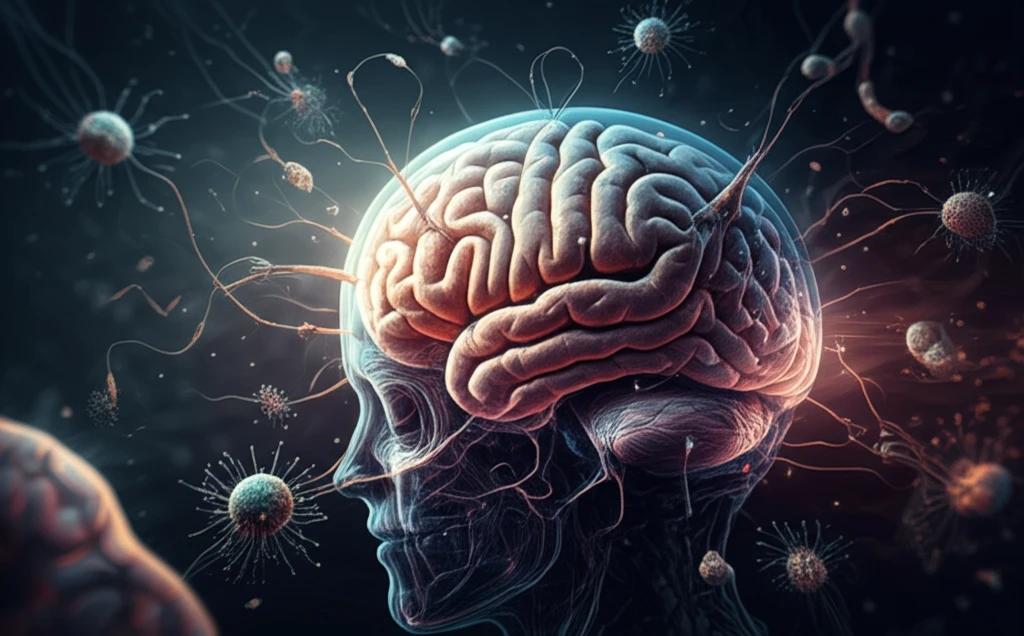
Imagine a microscopic world where tiny organisms can breach the body's most fortified defenses and invade the central nervous system. This is the reality of Acanthamoeba, a free-living amoeba capable of causing severe infections in humans. While often associated with eye infections and skin ulcerations, Acanthamoeba can also penetrate the brain, leading to a condition known as granulomatous amoebic encephalitis (GAE). This chronic infection, often mistaken for bacterial or viral invasions, is characterized by non-specific symptoms and a grim mortality rate exceeding 95%.
Acanthamoeba's journey to the brain typically begins elsewhere in the body, such as the cornea, skin, or lungs. From these sites, the amoebae can enter the bloodstream and eventually cross the blood-brain barrier (BBB), the protective shield that separates the brain from the circulatory system. Once inside the CNS, Acanthamoeba triggers a cascade of inflammatory responses, with neutrophils and macrophages releasing mediators that further compromise the BBB.
But what exactly happens inside the brain during an Acanthamoeba infection? Scientists are now focusing on the role of matrix metalloproteinases (MMPs), a family of enzymes involved in the breakdown of the extracellular matrix, the structural network surrounding brain cells. A recent study published in the International Journal of Molecular Sciences sheds light on the activity of MMPs and their tissue inhibitors (TIMPs) in the cerebral cortex and hippocampus during experimental Acanthamoeba infections.
The Brain's Response: MMPs and TIMPs in Action
The study explored how Acanthamoeba infections affect the levels of MMPs (specifically MMP-2 and MMP-9) and their corresponding TIMPs (TIMP-1 and TIMP-3) in the cerebral cortex and hippocampus, two critical brain regions involved in higher-level cognitive processing and memory. Researchers were particularly interested in the balance between MMPs and TIMPs, as an imbalance can lead to progressive pathological changes in the nervous system.
- MMP-2: Highest levels in the cerebral cortex 8 days post-infection in immunocompetent mice. Significantly higher levels in immunosuppressed mice at 16 days post-infection.
- MMP-9: Highest levels in the hippocampus of immunocompetent mice at 8 days post-infection. Significant increase in the cerebral cortex of immunocompetent mice at 8 days. Downregulation observed at 16 days, with a significantly lower level in the hippocampus of immunosuppressed mice at 24 days.
- TIMP-1: Significantly higher in the cerebral cortex of immunocompetent mice at 8 days post-infection. Downregulation in the hippocampus of immunocompetent mice compared to immunosuppressed mice at 8 days.
- TIMP-3: Significantly higher in the cerebral cortex of immunocompetent mice compared to immunosuppressed mice at 8 days post-infection. Downregulation in the hippocampus of immunosuppressed mice at 16 days in immunosuppressed mice.
Future Directions: Targeting MMPs for Brain Protection
The study underscores the potential of MMPs as therapeutic targets for limiting amoebic encephalitis. By understanding the specific roles of MMPs and TIMPs in the pathogenesis of Acanthamoeba brain infections, researchers hope to develop strategies to prevent the breaching of the blood-brain barrier and reduce nerve tissue damage. Further research is needed to fully characterize the complex interactions between the immune system, MMP activity, and neurodegeneration in the context of Acanthamoeba infections. The findings of this study offer a valuable foundation for future investigations aimed at developing effective treatments for this devastating condition.