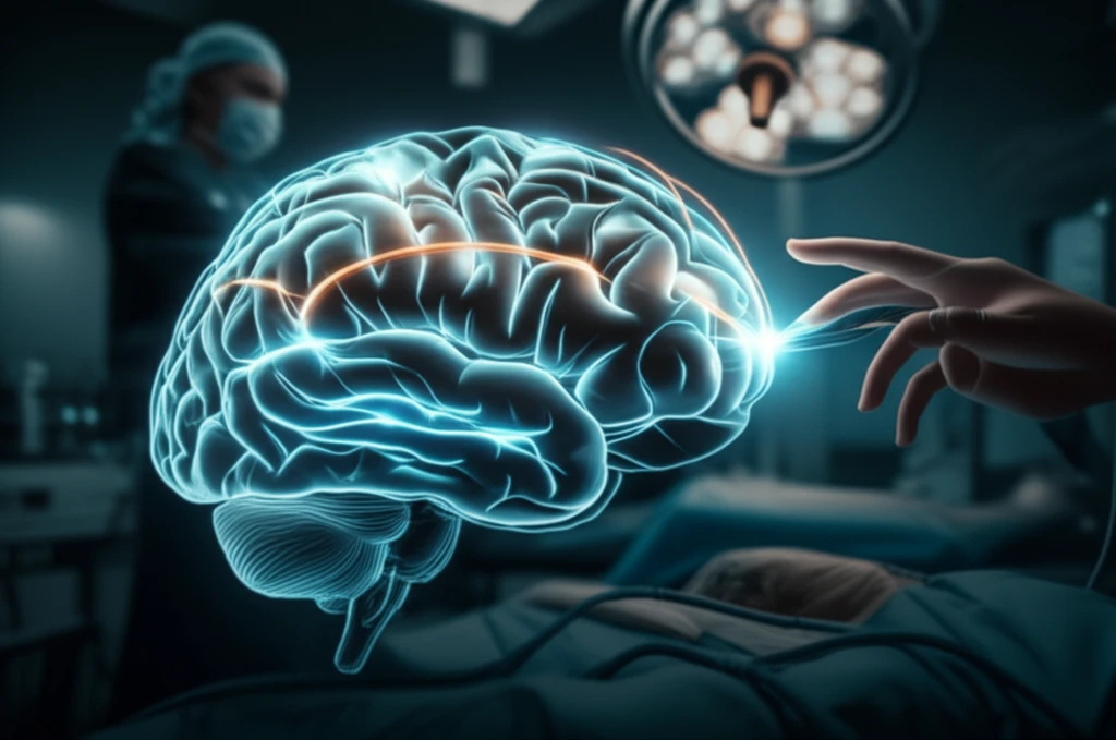
Brain Mapping Breakthrough: Minimizing Risks in Epilepsy Surgery
"Innovative neurophysiological monitoring refines surgical precision for congenital hemiparesis patients, promising safer outcomes and better functionality."
Epilepsy surgery presents a significant opportunity for individuals with drug-resistant seizures, especially those with congenital hemiparesis. However, the challenge lies in accurately targeting the seizure-causing areas while preserving essential brain functions. Traditional methods of brain mapping can be limiting, particularly in patients with developmental delays or cognitive impairments, underscoring the need for innovative approaches.
A recent study introduces a groundbreaking intraoperative neurophysiological monitoring (IONM) technique that refines the precision of epilepsy surgery. This method focuses on mapping corticospinal projections—the pathways that control movement—to minimize the risk of post-operative deficits. By employing a comprehensive approach to brain mapping, surgeons can make more informed decisions, improving patient outcomes and quality of life.
This article will delve into the specifics of this pioneering IONM technique, illustrating its application in a 34-year-old patient with congenital hemiparesis and intractable epilepsy. We'll explore how this method not only aids in surgical decision-making but also offers a promising alternative to conventional preoperative assessments.
Innovative Brain Mapping: A Step-by-Step Guide
The core of this new technique lies in its ability to map corticospinal projections in real-time during surgery. Unlike conventional methods, this IONM approach uses transcranial stimulation (TCS) to record compound muscle action potentials (CMAPs) from bilateral limb muscles simultaneously. This provides a comprehensive view of motor pathways, accounting for any atypical arrangements due to congenital conditions.
- Pre-Craniotomy TCS: Stimulating electrodes are placed strategically on the scalp (Cz, C3, and C4) to activate motor pathways.
- Bilateral CMAP Recording: Muscle responses are recorded from both sides of the body, revealing how each hemisphere contributes to motor function.
- Direct Cortical Stimulation (DCS): After opening the skull, surgeons use a monopolar electrode to directly stimulate the brain, further refining the map of motor areas.
- Integration and Resection: By combining TCS and DCS data, surgeons can precisely identify and preserve eloquent cortex while resecting the epileptogenic zone.
The Future of Safer Epilepsy Surgery
This innovative IONM technique represents a significant advancement in epilepsy surgery, particularly for patients with congenital hemiparesis. Its ability to map motor pathways in real-time, accounting for individual variations, minimizes the risk of post-operative deficits and maximizes the potential for successful seizure control.
Moreover, this method holds promise as a replacement for conventional preoperative assessments like functional MRI and transcranial magnetic stimulation, especially in young patients who may struggle with cooperation and participation. The simplicity and patient-independent nature of IONM make it a valuable tool for surgical planning and decision-making.
As research continues, this technique could become a standard of care, offering safer and more effective epilepsy surgery for a wider range of patients. The ability to predict functional outcomes and preserve quality of life will undoubtedly transform the landscape of epilepsy treatment.