
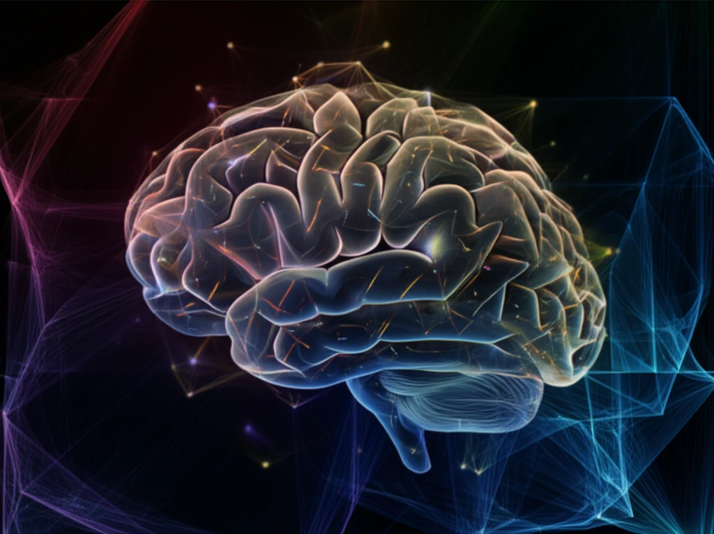
Decoding the Brain: How Cutting-Edge Tech is Mapping Our Thoughts

"Scientists are using advanced techniques to peer into the mind, but what are the implications for the future of mental health and understanding ourselves?"
The human brain, a complex network of billions of neurons, has long been an enigma. But now, thanks to rapidly evolving technologies, scientists are gaining unprecedented insights into its inner workings. Brain mapping, the process of creating detailed visual representations of the brain, is at the forefront of this revolution, offering the potential to unlock the secrets of our thoughts, emotions, and behaviors.
This article delves into the fascinating world of brain mapping, exploring the innovative techniques used to decode the brain's intricate architecture. From functional magnetic resonance imaging (fMRI) to independent component analysis (ICA), we'll examine how these tools are being used to create detailed maps of brain activity, with a specific focus on applications for mental health and cognitive enhancement.
We'll also explore the implications of these advancements, discussing how brain mapping could revolutionize the diagnosis and treatment of mental illnesses, while also raising important ethical considerations about the use of this powerful technology.
Mapping at Scale
NeuroQuery shows that building a global view of brain organization requires connecting evidence across widely different mental processes and mechanisms. The Reproducible Brain Charts resource extends this integrative approach by combining five large youth brain-development studies from three continents, covering 6,346 participants. Its harmonized psychiatric phenotypes use bifactor models to capture major dimensions of psychopathology.
Methods and Limits
Brain mapping generally combines imaging or electrophysiological measurements with computational analysis to relate brain properties to spatial representations. These methods can reveal patterns associated with structure or function, but they do not provide a complete or direct readout of mental activity. Interpretation is therefore shaped by measurement resolution, experimental design, analysis choices, and the limits of linking observed signals to cognition.
From Structure to Function
Human brain mapping has been pursued in various forms since antiquity, with modern maps evolving from structural descriptions toward functional and neuropsychological accounts. The field as presently known emerged when cognitive psychology’s experimental strategies were combined with brain-imaging techniques, first positron emission tomography and then functional magnetic resonance imaging. This combination helped researchers examine how brain function supports mental activities.
Unveiling the Mind: The Science Behind Brain Mapping
Brain mapping is not a single technique but a diverse field encompassing a range of methods, each offering unique insights into the brain's structure and function. One of the most prominent is fMRI, which measures brain activity by detecting changes associated with blood flow. By tracking these changes, scientists can create detailed maps of which brain regions are active during specific tasks or in response to various stimuli.
- fMRI (Functional Magnetic Resonance Imaging): Detects changes in blood flow to map brain activity.
- ICA (Independent Component Analysis): Identifies independent networks of brain activity.
- EEG (Electroencephalography): Measures electrical activity in the brain using electrodes.
- MEG (Magnetoencephalography): Detects magnetic fields produced by electrical activity.
Higher-Resolution Mapping
Recent brain-mapping research is expanding beyond conventional anatomical descriptions through advanced imaging and computational analysis. The cytoarchitecture-based link estimation, or CABLE, framework is presented as a way to enable three-dimensional mapping of brain fiber tracts while addressing limitations in accurate fiber reconstruction. Current reviews describe the field as multidisciplinary and focused on clarifying both structural and functional brain organization.
Resolution and Reliability
Brain-mapping results can be limited by both methodological assumptions and measurement scale. A reported challenge to lesion network mapping argues that this technique, used to link brain circuits with psychiatric disorders, neurological disease, and political behavior, may contain a serious flaw. Separately, fMRI voxels can contain millions of neurons and billions of synapses, making them relatively blunt instruments for analyzing micro-circuitry.
Comparing Mapping Approaches
Brain-mapping approaches differ in what they measure, the spatial or functional scale they can represent, and the assumptions required to interpret their outputs. Imaging methods can emphasize anatomy or activity, while electrophysiological approaches focus on electrical signals and their spatial or frequency patterns. Comparisons should therefore treat maps as complementary measurements rather than interchangeable pictures of thought.
The Future of the Mind: Applications and Ethical Considerations
Brain mapping holds tremendous promise for the future, particularly in the fields of mental health and cognitive enhancement. These new methods could revolutionize the diagnosis and treatment of mental illnesses, offering more personalized and effective interventions. However, with such powerful tools comes the need for careful ethical consideration. As we delve deeper into the mysteries of the brain, it's crucial to ensure that this technology is used responsibly and ethically, for the benefit of all.
Interpreting Brain Maps
Brain mapping is best understood as an evidence-building process rather than a literal translation of thoughts into pictures. Different techniques illuminate different properties of brain organization, and conclusions depend on how measurements are analyzed and related to mental functions. A careful synthesis should therefore distinguish observed neural patterns from broader claims about subjective experience.
Adaptive and AI-Assisted Mapping
One emerging direction is sensing-enabled adaptive neuromodulation, including BrainSense-enabled closed-loop deep brain stimulation that detects brain signals in real time to personalize stimulation therapy. Another is the use of AI to segment, align, fuse, and interpret MRI, fMRI, electron microscopy, transcriptomic, and connectomic data. The described 2026 workflows emphasize quality control, ground truth, and human review rather than relying on a single model.
The High-Resolution Challenge
Mapping the brain at cellular and sub-cellular resolution creates substantial data-driven modeling challenges. Reviews identify difficulties in building models of brain structure from high-resolution imaging datasets and discuss existing efforts and potential solutions. These challenges arise across both cellular and sub-cellular scales, making improved data strategies central to future progress.
People Behind the Maps
The practical meaning of brain mapping depends on how researchers, clinicians, and affected individuals interpret and use its results. Maps can support understanding of brain organization, but their implications should be communicated with care because measured patterns are not identical to personal thoughts or experiences. Human judgment remains important when translating technical findings into decisions about health, education, or society.