
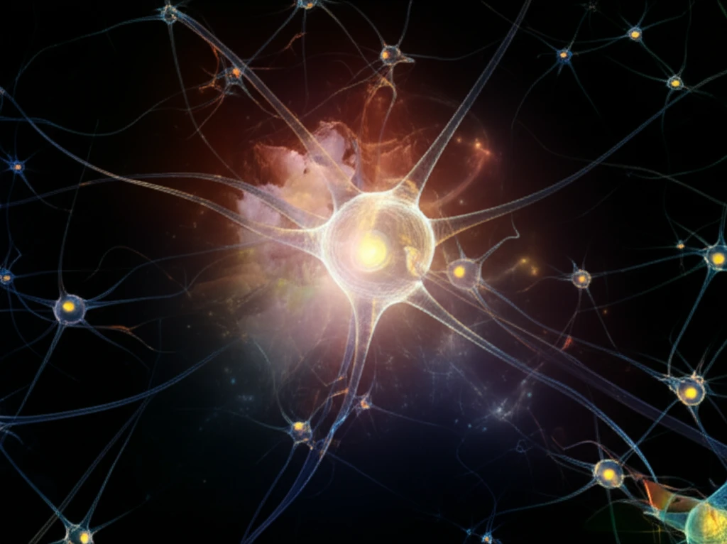
Brain Mapping: Unlocking Neuron Secrets for Healthier Minds

"New research maps individual neurons, revealing insights into brain function and potential treatments for neurological disorders."
The human brain, with its billions of interconnected neurons, remains one of the most complex and mysterious structures in the known universe. Understanding how individual neurons contribute to overall brain function is crucial for unraveling the mysteries of neurological disorders and developing effective treatments.
Traditional methods of studying the brain often provide a broad overview, failing to capture the intricate details of individual neurons and their specific roles. However, recent advances in neurogenomics are revolutionizing our ability to map the brain at an unprecedented level of detail.
A groundbreaking study published in Science details a novel approach that combines single-cell RNA sequencing and MERFISH (multiplexed error-robust fluorescence in situ hybridization) to map the identity and location of specific cell types within the mouse preoptic hypothalamus. This research provides an unbiased description of cell types, linking them to behaviors like sleep, thermoregulation, thirst, and social interaction.
Open Brain-Mapping Resources
The BrainMap website makes its software and data available free for educational and scientific, non-commercial use. Its coding scheme and taxonomy of experimental design are available without restriction. NeuroVault is a public repository for unthresholded statistical maps, brain parcellations, and atlases.
Methods and Mapping Challenges
Lesion network mapping (LNM), and atrophy network mapping for neurodegenerative brain clusters, have become widely adopted tools for linking focal lesions or clusters to distributed networks, according to a 2026 review. High-resolution brain mapping methods and computational approaches are advancing, but data-driven strategies for mapping large volumes still face challenges. A 2025 study describes a cytoarchitecture-based link estimation (CABLE) framework for 3D mapping of brain fiber tracts, addressing limitations in accurate fiber reconstruction.
From Structural to Functional Maps
A historical review traces human brain maps from ancient times to the present and describes a shift from purely structural mapping, through purely functional mapping, toward maps relating structure and function. Human functional brain mapping as it is known today emerged when cognitive psychology’s experimental strategies were combined with modern imaging. The review identifies positron emission tomography first, followed by functional magnetic resonance imaging, as techniques used to examine how brain function supports mental activities.
Decoding the Neuron Map: What Does It Reveal?
The study by Moffitt et al. represents a significant leap forward in our ability to understand the brain's cellular landscape. By mapping individual neurons and linking them to specific functions, researchers are gaining valuable insights into how the brain operates at its most fundamental level.
- Unprecedented Detail: Single-cell RNA sequencing allows researchers to analyze the genes expressed in individual neurons, providing a detailed molecular fingerprint of each cell.
- Spatial Mapping: MERFISH technology enables the precise localization of these neurons within specific brain regions, creating a spatial map of cell types.
- Functional Links: By correlating gene activity with specific behaviors, researchers can link cell types to their functions in processes like sleep, thermoregulation, thirst, and social interaction.
- Unbiased Approach: The methodology provides an unbiased description of cell types, reducing the reliance on prior assumptions and allowing for the discovery of novel cell populations.
Technical Limits in Spatial Mapping
Spatial transcriptomics methods such as seqFISH and MERFISH have improved the scalability of imaging-based approaches, but the methods can require lengthy sequential imaging and have small fields of view. The cited review also notes limits including lack of 3D resolution, high background signal, and constraints on gene selection from optical crowding or gene length. A separate review reports that current commercial platforms can profile only a few hundred to a thousand genes, citing optical crowding, increased binding-error risk across hybridization rounds, long imaging times, and small imaging areas. In an Alzheimer’s disease cell atlas, MERFISH gene panels failed to resolve a small number of non-neuronal types, likely because discriminant probes were lacking.
The Future of Brain Mapping: Implications and Applications
This research opens up exciting new avenues for understanding and treating neurological disorders. By identifying specific cell types and their roles in disease, researchers can develop targeted therapies that address the underlying causes of conditions like Alzheimer's disease, Parkinson's disease, and autism. The ability to map the brain at this level of detail also has implications for cognitive enhancement and personalized medicine, potentially leading to strategies for optimizing brain function and tailoring treatments to individual needs.
Integrating New Brain Maps
A 2025 review describes an emerging neuroscience paradigm that measures spatial patterns across multiple biological features to produce precise brain maps. It frames a key next question as how to integrate molecular, cellular, and dynamic features into a coherent understanding of brain structure and function. A neuroimaging overview points to quantum sensors and biophotonics as fields that may enable greater sensitivity, resolution, and non-invasive capabilities, while presenting these as prospective developments.
Challenges at Cellular Resolution
Brain mapping at cellular and sub-cellular resolutions presents challenges for developing data-driven strategies. A 2019 review highlights the difficulty of building models of brain structure from imaging datasets and discusses existing efforts to address those challenges and potential solutions. The field is multidisciplinary, combining advanced imaging techniques with computational analysis to study structural and functional organization.