Advanced MRI Techniques Reveal Microstructural Brain Damage in Traumatic Brain Injury

Brain Injury Breakthrough: New MRI Techniques Reveal Hidden Damage

"Advanced Diffusion MRI Uncovers Spatiotemporal Microstructural Changes After Traumatic Brain Injury, Opening New Doors for Diagnosis and Treatment"


Traumatic brain injury (TBI) is a global health concern, leading to significant disability and mortality. TBIs result from external forces, such as accidents, sports injuries, or violence, triggering complex cellular processes that can lead to long-term cognitive and behavioral impairments. While the impact of TBI on white matter has been extensively studied, the effects on gray matter have remained less understood, hindering comprehensive diagnosis and treatment strategies.

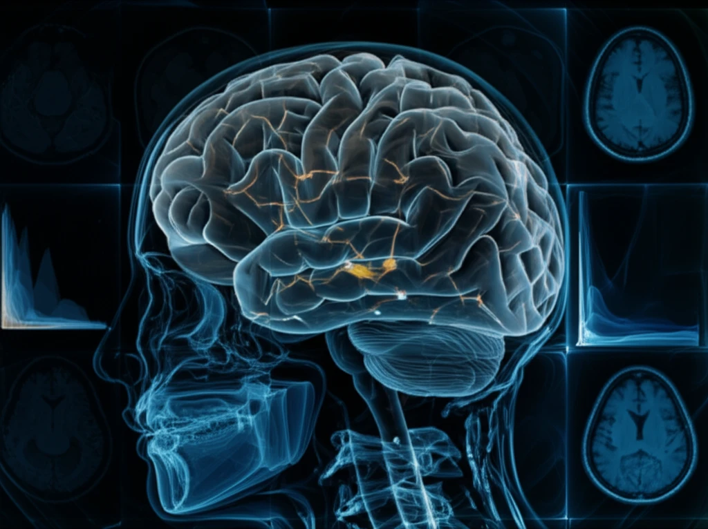
Magnetic Resonance Imaging (MRI) has become a crucial tool in diagnosing TBI-related changes in recent years. Traditional MRI scans, such as T1- and T2-weighted images, effectively assess primary lesions and edema. However, more advanced techniques like Diffusion Tensor Imaging (DTI) and Diffusion Kurtosis Imaging (DKI) are now being used to evaluate injury-associated changes at a microscopic level. DTI, based on a Gaussian model of water diffusion, helps examine white matter alterations, including axonal injury, demyelination, and edema.

A recent study published in the Journal of Neurotrauma sheds light on the potential of diffusion MRI to uncover spatiotemporal microstructural changes in gray matter following TBI. By combining DTI and DKI techniques with detailed histological analysis, researchers have provided a comprehensive view of the injury's progression in a rodent model, offering valuable insights for future clinical applications.

Unveiling Hidden Damage: How Diffusion MRI Works

Advanced MRI Techniques Reveal Microstructural Brain Damage in Traumatic Brain Injury

The study employed a controlled cortical impact (CCI) mouse model of TBI to investigate gray matter changes over time. Mice were scanned using T2-weighted structural imaging, DTI, and DKI at various intervals post-injury (5 hours, 1, 3, 7, 14, and 30 days). The MRI data was then validated with cross-sectional histopathology using cresyl violet staining and Glial Fibrillary Acidic Protein (GFAP) immunohistochemistry. This multimodal approach allowed researchers to correlate MRI findings with cellular-level changes, providing a comprehensive picture of TBI progression.

The results revealed a complex interplay of microstructural changes in both the ipsilateral (injured) and contralateral (opposite) hemispheres. Here are some of the key findings:

  • Lesion Volume: Lesion volume increased significantly up to 3 days post-injury, followed by a gradual decrease over the next month.
  • GFAP Signals: GFAP signals, indicating astrogliosis (the proliferation of astrocytes in response to injury), peaked on day 7 and persisted until day 30 in the ipsilateral and contralateral hippocampus, ipsilateral cortex, and thalamic areas.
  • Fractional Anisotropy (FA): FA, a measure of white matter integrity, increased in the pericontusional area on day 7 but decreased in the contralateral cortex, hippocampus, and thalamus.
  • Mean Diffusivity (MD): MD, reflecting water diffusion, was significantly lower in the pericontusional cortex. Increased MD and decreased mean kurtosis (MK) were limited to the injury site between days 7 and 30 and the contralateral hippocampus and thalamus on days 3 and 7.
The study demonstrates that DTI and DKI measures are sensitive to both ipsilateral and contralateral changes following TBI. These changes, particularly in FA, MD, and MK, reflect a complex interplay of cellular processes, including edema, inflammation, and astrogliosis. The findings highlight the potential of diffusion MRI to provide a more nuanced understanding of TBI pathology compared to traditional imaging techniques.

Why These Findings Matter

This research offers valuable insights into the complex microstructural changes that occur in the brain following TBI. By demonstrating the sensitivity of diffusion MRI to both ipsilateral and contralateral changes, the study opens new avenues for improved diagnostics and targeted treatments. Understanding the spatiotemporal dynamics of these changes could lead to earlier and more accurate identification of brain damage, ultimately improving patient outcomes. This study also highlights the potential of MRI techniques to track gliosis, a key marker of brain injury, in real time, offering new ways to monitor the effectiveness of therapies and interventions.

About this Article -

This article was crafted using a human-AI hybrid and collaborative approach. AI assisted our team with initial drafting, research insights, identifying key questions, and image generation. Our human editors guided topic selection, defined the angle, structured the content, ensured factual accuracy and relevance, refined the tone, and conducted thorough editing to deliver helpful, high-quality information.See our About page for more information.

Everything You Need To Know

1

What is a Traumatic Brain Injury (TBI) and why is it important to study?

Traumatic Brain Injury (TBI) results from external forces that trigger complex cellular processes, potentially leading to long-term cognitive and behavioral impairments. These injuries have traditionally been challenging to fully assess, especially in gray matter, which has been less understood compared to white matter injuries. Advanced Magnetic Resonance Imaging (MRI) techniques, like Diffusion Tensor Imaging (DTI) and Diffusion Kurtosis Imaging (DKI), are emerging as crucial tools because they can reveal microstructural changes associated with TBI. The study of TBI is critical due to its global impact on disability and mortality.

2

Why is Diffusion MRI important in understanding brain injuries?

Diffusion MRI is significant because it can uncover hidden damage at a microstructural level following a Traumatic Brain Injury (TBI). Traditional MRI scans, such as T1- and T2-weighted images, are useful for assessing primary lesions and edema, but diffusion MRI techniques like Diffusion Tensor Imaging (DTI) and Diffusion Kurtosis Imaging (DKI) provide a more detailed view of injury-associated changes. By assessing metrics like Fractional Anisotropy (FA), Mean Diffusivity (MD), and Mean Kurtosis (MK), diffusion MRI can reveal edema, inflammation, and astrogliosis, offering a more comprehensive understanding of TBI pathology. Diffusion MRI's sensitivity to both ipsilateral and contralateral changes, not detectable with standard methods, enables improved diagnostics and treatment strategies.

3

What are Diffusion Tensor Imaging (DTI) and Diffusion Kurtosis Imaging (DKI)?

Diffusion Tensor Imaging (DTI) is an MRI technique that examines white matter alterations resulting from injury. It utilizes a Gaussian model of water diffusion to assess changes such as axonal injury, demyelination, and edema. Fractional Anisotropy (FA), a metric derived from DTI, measures the integrity of white matter. Diffusion Kurtosis Imaging (DKI) is an advanced MRI technique that goes beyond DTI by providing additional information about the non-Gaussian diffusion of water in brain tissue. DKI helps assess changes in both white and gray matter, offering insights into the complexity of tissue microstructure following Traumatic Brain Injury (TBI). Mean Kurtosis (MK) is a key metric derived from DKI and can reflect changes in tissue microstructure such as edema and inflammation.

4

How was the study conducted to understand the gray matter changes after Traumatic Brain Injury (TBI)?

The study used a controlled cortical impact (CCI) mouse model to mimic Traumatic Brain Injury (TBI). The mice underwent MRI scans including T2-weighted structural imaging, Diffusion Tensor Imaging (DTI), and Diffusion Kurtosis Imaging (DKI) at various time points post-injury. Histological analysis, using cresyl violet staining and Glial Fibrillary Acidic Protein (GFAP) immunohistochemistry, was performed to validate the MRI findings by examining cellular-level changes. GFAP signals indicate astrogliosis, which is the proliferation of astrocytes in response to injury. This combined approach allowed researchers to correlate MRI data with cellular-level changes, creating a comprehensive understanding of how TBI progresses over time. Combining imaging and cellular analysis offers a robust way to validate and interpret the complex changes after TBI.

5

What do metrics like Fractional Anisotropy (FA) and Mean Diffusivity (MD) tell us about the brain after an injury?

Fractional Anisotropy (FA) is a measure of white matter integrity derived from Diffusion Tensor Imaging (DTI). In the study, FA increased in the pericontusional area on day 7 but decreased in the contralateral cortex, hippocampus, and thalamus. Mean Diffusivity (MD) reflects water diffusion and was found to be significantly lower in the pericontusional cortex. These changes, along with alterations in Mean Kurtosis (MK), indicate complex cellular processes such as edema, inflammation, and astrogliosis following Traumatic Brain Injury (TBI). The fact that these metrics change differently in various brain regions at different times shows the intricate spatiotemporal dynamics of TBI pathology.

Newsletter Subscribe

Subscribe to get the latest articles and insights directly in your inbox.