Brain Breakthrough: How Tiny Microscopes Are Revolutionizing Neuroscience and Mental Health
"Unlocking the Secrets of the Brain: New Techniques Improve Brain Imaging, Paving the Way for Better Mental Health Treatments"
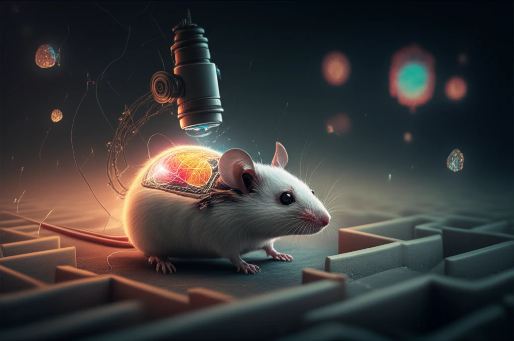
Imagine a world where scientists can watch brain activity in real-time, like a movie, as thoughts and emotions unfold. This isn't science fiction; it's the reality of modern neuroscience, thanks to groundbreaking advancements in microscopy. Scientists are now using tiny, head-mounted microscopes to observe the activity of individual neurons in living, moving animals. This leap in technology is providing unprecedented insights into the complexities of the brain, potentially leading to transformative treatments for mental health disorders and neurological conditions.
The key to this revolution lies in the development of miniature microscopes that are small and light enough to be worn by freely moving animals, such as mice. These innovative devices allow researchers to study brain activity in natural settings, providing a more accurate picture of how the brain functions. However, getting clear, detailed images of brain activity in these conditions has been a significant challenge, until now. New surgical techniques are further improving the clarity of images, opening new possibilities for research.
This article will explore the latest advancements in brain imaging, focusing on the use of miniature microscopes and innovative surgical techniques. We'll delve into the challenges researchers have faced and how they're overcoming them. By understanding these advancements, we can better appreciate the profound impact they're having on our understanding of the brain and the potential for improved mental health treatments. We'll also highlight the specific research that's paving the way for these exciting breakthroughs.
The Challenges of Brain Imaging: Why Clear Images Matter
Getting a clear view of the brain isn't as simple as it sounds. Traditional brain imaging techniques have limitations. For example, traditional open-skull cranial window surgery can lead to inflammation and tissue overgrowth, which clouds the images. The brain is a delicate organ, and any surgical procedure can cause damage, affecting the quality of the images and the health of the brain tissue itself. This makes it difficult to study brain activity over extended periods, hindering the ability to observe long-term changes and responses to treatments.
- Inflammation and Tissue Overgrowth: Traditional methods often lead to clouding of images.
- Minimizing Brain Damage: Reducing harm is essential for accurate imaging.
- Need for Natural Behavior: Animals need to move freely for realistic study.
- Positioning and Focus: Precise microscope placement is critical.
The Future of Neuroscience: A Brighter Outlook for Mental Health
The advancements in brain imaging technology represent a significant step forward in neuroscience. The ability to study brain activity with greater clarity and precision opens doors to new discoveries. This progress offers a brighter outlook for understanding and treating mental health disorders and neurological conditions. As research continues, we can expect even more exciting breakthroughs, offering hope for a healthier future for all. The potential for more effective treatments, earlier diagnoses, and a deeper understanding of the human mind is within reach, thanks to the innovative work of scientists around the world. The future of mental health looks brighter than ever before.