Surreal illustration of brachial plexus nerve network with MRI scan lines.

Brachial Plexus Injuries: Can MRI Scans Accurately Detect Nerve Damage?

"A close look at the effectiveness of 1.5T MRI in diagnosing root avulsions after traumatic injuries."


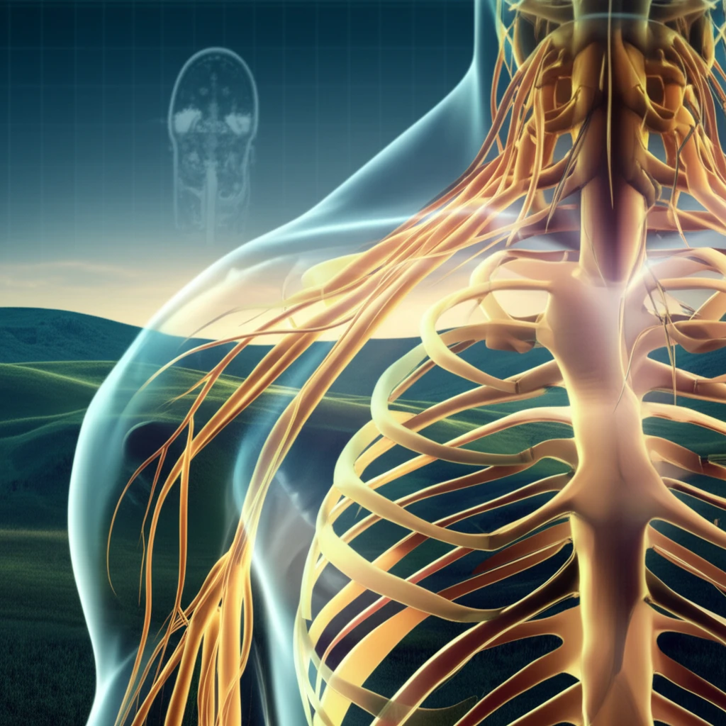
Traumatic brachial plexus injuries, which affect up to 1% of adults involved in serious road accidents, can have devastating consequences. These injuries involve damage to the network of nerves that control movement and sensation in the arm and hand. Effective treatment and recovery depend heavily on accurately diagnosing whether the nerve roots have been avulsed, or torn away, from the spinal cord.

Distinguishing between pre-ganglionic injuries (root avulsions) and post-ganglionic injuries (ruptures or attenuations) is crucial because each type requires a different surgical approach and has a different prognosis. Post-ganglionic injuries, where the nerve is still attached to the spinal cord, can often be repaired or grafted, offering a more favorable outcome. Root avulsions, on the other hand, typically require nerve transfers from other areas of the body, as re-implantation of the avulsed root remains uncertain.

Magnetic resonance imaging (MRI) has become increasingly popular for evaluating brachial plexus injuries due to its ability to provide detailed images of soft tissues. However, the accuracy of MRI in detecting root avulsions has been a topic of debate. This article explores the diagnostic accuracy of 1.5T MRI in detecting root avulsions in adults with traumatic brachial plexus injuries, shedding light on the reliability and limitations of this imaging technique.

What is the Role of MRI in Diagnosing Brachial Plexus Injuries?

Surreal illustration of brachial plexus nerve network with MRI scan lines.

Currently, the most reliable method for identifying root avulsions involves surgical exploration of the supraclavicular brachial plexus, where surgeons directly visualize the nerves. Given the invasive nature and uncertain outcomes of exploratory surgery, doctors often rely on pre-operative imaging and neurophysiological tests to inform their surgical decisions. MRI is valued for its multi-planar capabilities and superior soft-tissue contrast, allowing doctors to visualize the nerve roots and surrounding structures in detail.

Despite its advantages, the diagnostic accuracy of MRI for root avulsions has been inconsistent. Previous studies have reported varying accuracy rates, ranging from 52% to 88%. Technical limitations and the use of clinical follow-up or electrophysiological studies as reference standards have contributed to these inconsistencies. To address these limitations, a new study has investigated the diagnostic accuracy of 1.5T MRI, using operative exploration as the reference standard.

  • Study Design: A retrospective cohort study was conducted, evaluating MRI scans performed on a consecutive series of adult males with non-penetrating traumatic brachial plexus injuries.
  • Participants: The study included patients managed at a single institution between January 2008 and July 2016.
  • Index Test: MRI of the brachial plexus was performed using a 1.5 Tesla scanner (Siemens Avanto). Specific sequences included sagittal T1-weighted and T2-weighted turbo-spin echo, axial T2 turbo-spin echo (TSE), coronal short-tau inversion recovery (STIR), and constructive interference steady state (CISS).
  • Reference Standard: Operative exploration of the supraclavicular brachial plexus was used as the reference standard for diagnosing root avulsions. Avulsion was defined based on the absence of nerve roots in the foramina or the ease with which neural structures could be pulled away during exploration.
  • Data Analysis: Statistical methods were used to assess the diagnostic accuracy of MRI, including sensitivity, specificity, positive predictive value, and negative predictive value.
The study found that the diagnostic accuracy of MRI for detecting root avulsions of C5-T1 was 79%. The diagnostic accuracy of pseudomeningoceles, which are expansions of the space containing the nerve root and cerebrospinal fluid, as a surrogate marker of root avulsions was 68%. The study concluded that pseudomeningoceles are not a reliable sign of root avulsion and that MRI has modest diagnostic accuracy for root avulsions in the context of adult traumatic brachial plexus injuries.

Key Takeaways and Future Directions

The study highlights the importance of improving diagnostic imaging techniques for brachial plexus injuries. While MRI is a valuable tool, its modest diagnostic accuracy underscores the need for continued research and development in this area. Future studies could explore the use of advanced MRI techniques, such as diffusion tensor imaging and hyperpolarization methods, to enhance the accuracy of root avulsion detection. Additionally, the study recommends that patients with brachial plexus injuries be promptly referred to specialist centers to facilitate high-quality prospective research and improve patient outcomes.

About this Article -

This article was crafted using a human-AI hybrid and collaborative approach. AI assisted our team with initial drafting, research insights, identifying key questions, and image generation. Our human editors guided topic selection, defined the angle, structured the content, ensured factual accuracy and relevance, refined the tone, and conducted thorough editing to deliver helpful, high-quality information.See our About page for more information.

Everything You Need To Know

1

What are brachial plexus injuries, and why is accurate diagnosis crucial?

Brachial plexus injuries involve damage to the network of nerves controlling arm and hand movement and sensation. Accurate diagnosis is critical because it determines the treatment approach and prognosis. Distinguishing between pre-ganglionic injuries, like root avulsions, and post-ganglionic injuries, such as ruptures, is essential. Root avulsions often require nerve transfers, while post-ganglionic injuries can sometimes be repaired or grafted, impacting the potential for recovery.

2

How is MRI used to diagnose brachial plexus injuries, and what are its limitations?

Magnetic Resonance Imaging (MRI) is used to evaluate brachial plexus injuries due to its ability to provide detailed images of soft tissues, including the nerve roots. A 1.5 Tesla scanner (Siemens Avanto) can visualize the nerve roots and surrounding structures. However, the diagnostic accuracy of MRI for detecting root avulsions has been inconsistent, with varying accuracy rates reported in previous studies. This study found that MRI has a modest diagnostic accuracy of 79% for root avulsions of C5-T1.

3

What is the significance of distinguishing between pre-ganglionic and post-ganglionic injuries in brachial plexus injuries?

The distinction between pre-ganglionic and post-ganglionic injuries is of utmost importance because it dictates the course of treatment and the expected outcome. Pre-ganglionic injuries, specifically root avulsions, involve the tearing of the nerve roots from the spinal cord. These injuries typically require nerve transfers from other areas of the body. Post-ganglionic injuries, where the nerve remains connected to the spinal cord, offer a better chance of recovery through repair or grafting.

4

What were the key findings of the study on 1.5T MRI for brachial plexus injuries?

The study, which used operative exploration as the reference standard, found that the diagnostic accuracy of 1.5T MRI for detecting root avulsions of C5-T1 was 79%. The study also examined the diagnostic accuracy of pseudomeningoceles as a surrogate marker for root avulsions, finding it to be 68%. The study concluded that pseudomeningoceles are not a reliable sign of root avulsion and that MRI has modest diagnostic accuracy for root avulsions in the context of adult traumatic brachial plexus injuries.

5

What are the implications of this research, and what future directions are suggested for improving the diagnosis of brachial plexus injuries?

The research highlights the need for improved diagnostic imaging techniques for brachial plexus injuries. While MRI is a valuable tool, its modest diagnostic accuracy underscores the need for further research. Future studies could explore advanced MRI techniques, such as diffusion tensor imaging and hyperpolarization methods, to enhance the accuracy of root avulsion detection. The study recommends prompt referral of patients with brachial plexus injuries to specialist centers to facilitate high-quality prospective research and improve patient outcomes.

Newsletter Subscribe

Subscribe to get the latest articles and insights directly in your inbox.