
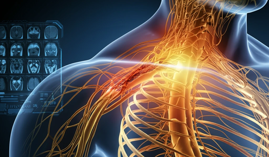
Brachial Plexus Injuries: Can MRI Scans Accurately Detect Nerve Root Damage?

"A new study examines the effectiveness of magnetic resonance imaging (MRI) in diagnosing root avulsions in adults following traumatic brachial plexus injuries."
Traumatic brachial plexus injuries can occur from traffic collisions or high-impact accidents and often affect adults, leading to significant disability. These injuries involve damage to the network of nerves (the brachial plexus) that controls movement and sensation in the arm and hand. The type and severity of the injury are crucial for determining the best course of treatment and predicting recovery.
Injuries are generally classified as either pre-ganglionic or post-ganglionic. Post-ganglionic injuries, such as nerve ruptures or attenuations, typically have a better prognosis because they can be repaired with surgery or nerve grafts. Pre-ganglionic injuries, also known as root avulsions, involve the nerve roots being torn away from the spinal cord. These injuries are more complex and often require nerve transfers from other parts of the body, as re-implantation of the avulsed root is often unsuccessful.
Identifying root avulsions is therefore critical because it significantly alters the surgical plan and the expected outcome. Doctors often use exploratory surgery to assess the damage, but imaging techniques like magnetic resonance imaging (MRI) are becoming increasingly popular for pre-operative assessment. MRI offers detailed images of the soft tissues in the brachial plexus, but questions remain about its accuracy in detecting root avulsions.
Current Impact
Brachial plexus injuries can affect sensation, movement, and strength in the shoulder, arm, forearm, and hand. Their impact varies according to which nerves are affected and how severely they are damaged. MRI may contribute to evaluation, but its ability to detect nerve root damage can depend on injury characteristics and clinical context.
Anatomy and Clinical Evaluation
The brachial plexus is a network formed by the anterior rami of the lower four cervical nerves and the first thoracic nerve, C5 through T1. It extends from the spinal cord through the neck, over the first rib, and into the armpit, supplying nerve fibers to the chest, shoulder, arm, forearm, and hand. The Mayo Clinic describes brachial plexus injuries as occurring when these nerves are stretched, compressed, ripped apart, or torn away from the spinal cord. Because the brachial plexus follows a complex anatomical course, assessing the location and severity of damage can be challenging and may require correlation between imaging findings and clinical examination.
Evolving Understanding
Understanding of brachial plexus injuries has developed alongside broader knowledge of nerve anatomy and neurological examination. Earlier approaches generally relied heavily on symptoms, physical examination, and anatomical localization. Modern imaging has added ways to examine structures that may not be directly visible clinically. The historical record suggests a gradual expansion of diagnostic tools rather than a single definitive milestone.
MRI Accuracy in Diagnosing Root Avulsions: What the Study Shows
A recent study published in the Journal of Hand Surgery (European Volume) investigated the accuracy of MRI in detecting root avulsions in adults with traumatic brachial plexus injuries. The study retrospectively analyzed data from 29 male patients who underwent both MRI and exploratory surgery at a single institution between 2008 and 2016. The goal was to determine how well MRI results matched the findings during surgery, which is considered the gold standard for diagnosis.
- Root Avulsion: Nerve roots torn away from the spinal cord.
- Pre-ganglionic Injury: Injury occurring before the nerve cell body.
- Post-ganglionic Injury: Injury occurring after the nerve cell body.
- Pseudomeningocele: Fluid-filled sac that forms when a nerve root is torn from the spinal cord.
- MRI (Magnetic Resonance Imaging): A detailed imaging technique using magnetic fields and radio waves to create images of the body's soft tissues.
Current Research
Recent research continues to examine how accurately MRI can characterize brachial plexus and nerve root injuries. Investigators are interested in improving visualization of injury location, severity, and associated structural changes. Findings may vary with MRI technique, timing, and the type of nerve damage being assessed. MRI is therefore best considered part of an evolving diagnostic field rather than an infallible standalone test.
Diagnostic Limitations
MRI does not necessarily provide a complete picture of nerve function or recovery potential. Structural abnormalities may be difficult to distinguish from clinically meaningful nerve root damage in some cases. Imaging can also be limited by the complexity of the brachial plexus and by changes that develop over time. These limitations support interpreting MRI alongside symptoms, examination findings, and other available assessments.
Comparing Diagnostic Tools
MRI offers a noninvasive way to evaluate the brachial plexus and nearby nerve roots. Other diagnostic approaches may provide complementary information about nerve function or the broader clinical pattern of injury. No single method necessarily answers every diagnostic question. The most useful comparison is therefore based on what each test can reveal, rather than assuming that one method is universally superior.
Implications for Patients and Future Research
While MRI is a valuable tool for assessing brachial plexus injuries, this study highlights its limitations in accurately diagnosing root avulsions. The findings suggest that doctors should not rely solely on MRI results when making treatment decisions. Surgical exploration, performed by an experienced surgeon, remains the most reliable method for determining the extent of the damage and planning the appropriate reconstructive procedures. Further research is needed to improve the accuracy of MRI and other imaging techniques for diagnosing brachial plexus injuries. This could involve using higher-resolution MRI scanners, advanced imaging sequences, or combining MRI with other diagnostic tests, such as diffusion tensor imaging or hyperpolarization methods. Ultimately, the goal is to provide patients with more accurate diagnoses and better treatment outcomes.
Putting MRI in Context
MRI can be valuable when clinicians need anatomical information about suspected brachial plexus or nerve root injury. Its findings should be interpreted in relation to the patient's symptoms and neurological examination. A scan may support a diagnosis without fully determining functional severity or prognosis. Overall, accurate assessment depends on integrating imaging with clinical judgment.
Future Directions
Future work may focus on making MRI more sensitive, more precise, and better suited to distinguishing different types of nerve injury. Researchers may also seek clearer links between imaging findings and patient outcomes. Improvements in imaging protocols and interpretation could strengthen its role in diagnosis. Even so, future advances are likely to complement rather than replace clinical assessment.
Wider Clinical Challenges
Brachial plexus injuries are difficult to evaluate because the plexus contains multiple nerve roots that travel through a complex anatomical region. Diagnostic decisions may also be affected by injury severity, timing, and the availability of specialized expertise. These factors can make consistent assessment challenging across clinical settings. Broader progress therefore depends on both better imaging and reliable integration of clinical information.
Patient Experience
Brachial plexus injuries can interfere with movement, sensation, and everyday use of the shoulder, arm, forearm, or hand. The consequences may vary widely depending on which nerves are affected and whether they are stretched, compressed, torn, or separated from the spinal cord. For patients, the practical meaning of an MRI result is therefore closely tied to symptoms and functional limitations. Clear communication about what imaging can and cannot show is an important part of care.