
Back Pain Breakthrough: Are Your Pedicle Screws Missing the Mark?

"New research suggests traditional spinal fusion techniques might be overlooking key areas for optimal bone density and fixation strength. Discover how alternative placements could revolutionize back pain treatment."
Chronic back pain is a widespread issue, and for many, spinal fusion surgery offers a beacon of hope. A critical factor in the success of these procedures is the strength of the bone where implants are anchored. Traditionally, surgeons have relied on pedicle screws, assuming the pedicles—small, paired bony structures that connect the vertebral body to the posterior elements—offer the best fixation points. But what if this assumption is wrong?
A groundbreaking study is challenging this long-held belief, suggesting that pedicles might not be the densest regions of the lumbar vertebrae. This could have significant implications for how spinal fusion surgeries are performed, especially for individuals with osteoporosis or other conditions that compromise bone quality. The research opens the door to exploring alternative sites that could provide more secure and lasting fixation, ultimately reducing the risk of implant failure and the need for revision surgeries.
The quest for more effective spinal fusion techniques is particularly crucial given the aging population and the increasing prevalence of osteoporosis. As more individuals undergo these procedures, understanding the nuances of bone density within the vertebrae becomes paramount. This article delves into the findings of this study and what it means for the future of back pain treatment.
Success Rates vs. Ten-Year Realities
Spinal fusion, also called spondylosyndesis or spondylodesis, is a surgery that connects two or more vertebrae to stabilize the spine and ease pain, and it is often recommended for issues such as degenerative disc disease and scoliosis. Utilization is rising sharply in the United States, with the number of lumbar fusions per year in New York State increasing by 55% from 2005 to 2014. Success rates often exceed 80% in many studies, reflecting improvements in surgery and postoperative care. Longer-term outcomes are more sobering, as reported success rates drop after 10 years, primarily due to reoperation and adjacent segment disease.
Graft Choices and the Limits of Newer Approaches
Posterior fusion approaches, performed through an incision in the back, remain a mainstay, with techniques selected according to the spinal level and condition. Autograft and allograft bone, the standard approaches for lumbar fusion, carry important disadvantages. Bone graft substitutes such as recombinant human bone morphogenetic proteins (rhBMP-2 and rhBMP-7) have emerged as viable alternatives. Anatomical limits constrain some newer techniques: the extreme lateral interbody fusion (XLIF) approach has shown good results but is generally limited at the lumbar spine to segments L1-L5, and a single-center report of three cases applied it at the L5-S1 level.
A Century of Fusing the Spine
Historical accounts trace spine fusion operations back through the 19th and 20th centuries, documenting the evolution of the procedure. For scoliosis in particular, surgical treatment has long involved extensive spinal fusions, at times spanning from the cervical spine to the pelvis. Long-term patient accounts offer a modern view of that legacy: one woman, six years out from her fusion at UVA Health, reports she could not be happier with her choice. Another patient traces a path that began with a 2003 motorcycle accident in Phoenix and led, years later, to needing fusion surgery.
The Bone Density Puzzle: Rethinking Spinal Fusion
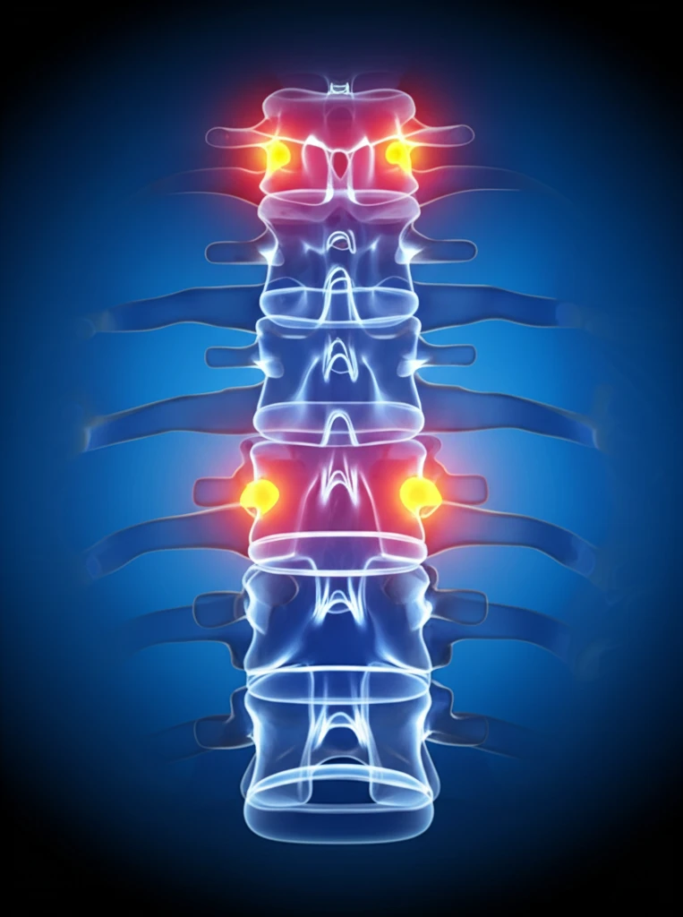
The study, a detailed examination of cadaveric lumbar vertebrae, employed computed tomography (CT) imaging to assess bone mineral density (BMD) across various anatomical subregions. Researchers digitally isolated the spine and meticulously segmented each vertebra into seven distinct regions: the vertebral body, pedicles, transverse processes, lamina, superior articular processes, inferior articular processes, and spinous process. By converting Hounsfield units (HU), a measure of radiodensity obtained from CT scans, into BMD values, the team was able to map the density landscape of each vertebra.
- The lamina and inferior articular processes have the highest bone mineral density in the lumbar vertebrae.
- The vertebral body has the lowest bone mineral density.
- Pedicles have moderate bone mineral density, lower than the lamina and inferior articular processes.
- These findings challenge the traditional focus on pedicles as the primary fixation point in spinal fusion surgery.
What the Evidence Shows
Spinal fusion is a highly invasive surgery in which an implant is placed in the spine to prevent movement between bones. A Monash University review of the evidence notes that lumbar spine fusion is a common procedure associated with a high cost burden and risk of serious complications, and it summarizes systematic reviews on the procedure's effectiveness across most diagnoses. Recovery timelines are substantial, with patients having a standard spinal fusion potentially taking six months or longer for a full recovery. Degenerative conditions of the lumbar spine, such as a herniated disc or spinal stenosis, are common causes of chronic back pain that lead patients to consider the surgery.
When Fusion Is Not the Only Answer
Spinal fusion is typically reserved for situations where conservative treatments have failed to provide adequate relief, or where there is instability or deformity in the lumbar spine. Even after surgery, patients must adapt everyday movement: one rehabilitation-focused tutorial demonstrates a modified sit-up technique using a small Pilates ball to challenge the abdominal muscles without forcing the spine into uncomfortable positions. Alternatives exist but do not always match the situation, since spinal decompression and inversion are distinct from cases where the spinal cord or nerves are squeezed by an injury or a diagnosed spinal problem. Case reports illustrate the complexity of surgical candidates, including a thoracic spine fusion involving an obese male patient with a multi-year history of lower leg pain and numbness.
Fusion vs. Disc Replacement vs. Graft Choice
Both spinal fusion and artificial disc replacement can reduce pain when the diagnosis is correct, and the key difference lies in what happens to movement at the treated level. Dr. Todd Lanman highlights the significant differences between the two procedures, which is an important consideration in patient counseling. Radiographic imaging plays a central role, since spinal fusion x-rays are essential for planning and checking surgery and show how the spine's alignment changes from before to after the procedure. Graft choice is also compared, with studies weighing fusion using the patient's own bone against the bone substitute ProOsteon 200 for neck fusion.
The Future of Spinal Fusion: A Shift in Perspective
This research signals a potential paradigm shift in how we approach spinal fusion surgery. By recognizing the variations in bone density within the vertebrae and exploring alternative fixation sites, surgeons can potentially improve outcomes, reduce complications, and enhance the quality of life for countless individuals suffering from chronic back pain. Further research is needed to validate these findings and refine surgical techniques, but the future of spinal fusion looks promising, with a renewed focus on personalized approaches and optimized implant placement.
What the Experts Advise
Expert guidance stresses that the decision for spinal fusion should come only after non-surgical alternatives have been exhausted, followed by a careful risk-benefit analysis with a specialist. Laminectomy and spinal fusion are two common surgeries that help with back pain and spine health, and expert surgical insight can help patients understand their options. Specialists also advise on procedure types, recovery expectations, and risks, alongside minimally invasive alternatives for eligible patients.
Materials, Minimally Invasive Care, and Personalized Medicine
Market outlooks for lumbar spine fusion point to several emerging trends: the introduction of advanced implant materials, rising demand for outpatient spine surgeries, and an increasing focus on personalized medicine in spine fusion procedures. Much of this innovation is driven by fusion's known complications, since spinal fusion can place pressure on the bones above and below the fused segment, causing them to break down and degenerate more rapidly. This adjacent segment disease increases the likelihood of future revision surgery, motivating less invasive and more targeted approaches. Long-term patient accounts, including those ten years out from surgery, provide real-world perspective on how these outcomes actually play out.
Smoking, Rehab, and the Support Network
Lifestyle factors weigh heavily on outcomes, since smoking creates a biologically challenging environment in which spinal fusion is less likely to succeed, and nicotine has been shown to have a direct negative effect on bone graft healing. Structured recovery matters as well, with rehabilitation protocols from leading orthopedic institutions compared on weight-bearing status, key restrictions, and phased progression. Patient communities describe fusion as a big deal that can provide great relief, while noting alternatives such as artificial discs. Individual patients emphasize that fusion does not mean a lifetime of pain, and that the right daily movements can make all the difference in staying strong, mobile, and pain-free.
Patients Behind the Procedures
A real-world study of minimally invasive posterior thoracic procedures demonstrated outcomes comparable to open surgery in length of stay, with added benefits of shorter procedure times and lower intraoperative blood loss. Individual cases illustrate the human stakes: one Mozambican patient endured 20 years of chronic back pain before advanced minimally invasive spine fusion restored his quality of life. That case exemplifies the remarkable impact that precise diagnosis, skilled surgical intervention, and dedicated patient involvement can have after prolonged suffering.