Surreal illustration of uranium worker fading into DNA, symbolizing radiation exposure and genetic damage.

Are We Underestimating the Risks? Genetic Damage in Uranium Processing Workers

"A new study analyzes genetic damage in former uranium processing workers, revealing surprising insights into radiation exposure and long-term health effects."


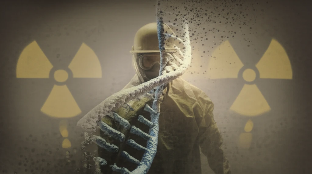
For years, the health risks associated with radiation exposure have been a significant concern, particularly for individuals working in industries dealing with radioactive materials. Uranium processing, an essential part of the nuclear fuel cycle, exposes workers to various forms of radiation, raising questions about the potential long-term effects on their health. Understanding the nature and extent of genetic damage in these workers is crucial for developing effective safety measures and healthcare strategies.

A recent study published in Cytogenetic and Genome Research delves into the analysis of genetic damage in lymphocytes—a type of white blood cell—of former uranium processing workers. The research focuses on individuals who were employed at the MAPE Mydlovary plant, a uranium processing facility in the Czech Republic that ceased operations in 1991. By examining the frequency of cells containing micronuclei (MN) and the presence of centromeres in these MN, the study aims to shed light on the subtle yet significant impacts of radiation on the workers' genetic material.

This research is particularly important because it challenges some of the existing assumptions about radiation exposure and its effects. While previous studies have focused on uranium miners, this investigation looks at processing workers, who have different exposure pathways. Understanding these differences can help refine our knowledge of how various forms of radiation affect human health and what specific precautions are needed in different industrial settings. The findings could potentially influence future radiation safety protocols and healthcare practices for workers in similar environments.

Decoding the Study: What Did Researchers Measure?

Surreal illustration of uranium worker fading into DNA, symbolizing radiation exposure and genetic damage.

The research team, led by Friedo Zölzer and colleagues, analyzed blood samples from 98 men in Southern Bohemia, Czech Republic. Of these, 46 had previously worked at the MAPE Mydlovary uranium processing plant, while 52 were controls from the same area. This setup allowed for a comparative analysis between those with known radiation exposure and those without, providing a baseline for understanding the specific impacts of uranium processing work.

The researchers used a sophisticated technique called fluorescence in situ hybridization (FISH) to detect micronuclei in the lymphocytes. Micronuclei are small, additional nuclei that form in cells when chromosomes or chromosome fragments are not properly included during cell division. The presence of micronuclei can indicate genetic instability and damage, making them a useful marker for radiation exposure. The FISH technique allowed the team to determine whether these micronuclei contained centromeres (CEN+), indicating that a whole chromosome was involved, or were centromere-negative (CEN-), suggesting chromosome fragments were the cause.

  • Collection of Blood Samples: Blood samples were collected from 98 male individuals, 46 of whom were former workers at the MAPE Mydlovary uranium processing plant, and 52 controls from the same area.
  • FISH Technique: Fluorescence in situ hybridization (FISH) was used to detect micronuclei (MN) in lymphocytes, identifying those with centromeres (CEN+) and those without (CEN-).
  • Analysis of Binucleated Cells (BNC): A total of 1,000 binucleated cells (BNC) per participant were analyzed after cytochalasin B treatment to assess genetic damage.
  • Statistical Analysis: Statistical methods, including Student's t-test, were used to compare the data between the exposed and control groups.
To ensure accurate results, the team analyzed 1,000 binucleated cells (BNC) per participant after treating them with cytochalasin B, a chemical that prevents cell division but allows the nucleus to divide, making it easier to identify micronuclei. The findings were then subjected to rigorous statistical analysis to determine if there were any significant differences between the exposed workers and the control group. This comprehensive approach helped to ensure the reliability and validity of the study’s conclusions.

The Bigger Picture: Implications and Future Research

This study provides a valuable contribution to our understanding of the long-term health effects of radiation exposure in uranium processing workers. While the results may seem surprising, they highlight the complexity of the relationship between radiation and genetic damage. Future research should focus on further exploring the role of radon exposure and genomic instability, as well as investigating the potential impact of other environmental factors. Ultimately, this knowledge will help in refining radiation safety protocols and developing more effective healthcare strategies for those who work in potentially hazardous environments.

About this Article -

This article was crafted using a human-AI hybrid and collaborative approach. AI assisted our team with initial drafting, research insights, identifying key questions, and image generation. Our human editors guided topic selection, defined the angle, structured the content, ensured factual accuracy and relevance, refined the tone, and conducted thorough editing to deliver helpful, high-quality information.See our About page for more information.

This article is based on research published under:

DOI-LINK: 10.1159/000441889, Alternate LINK

Title: Analysis Of Genetic Damage In Lymphocytes Of Former Uranium Processing Workers

Subject: Genetics (clinical)

Journal: Cytogenetic and Genome Research

Publisher: S. Karger AG

Authors: Friedo Zölzer, Renata Havránková, Zuzana Freitinger Skalická, Andrea Rössnerová, Radim J. Šrám

Published: 2015-01-01

Everything You Need To Know

1

What specific cells were examined to assess genetic damage in the study?

The study analyzed genetic damage in former uranium processing workers by examining their lymphocytes, a type of white blood cell. These cells were collected from individuals who worked at the MAPE Mydlovary uranium processing plant. The researchers specifically looked for micronuclei (MN) within these cells, using a technique called fluorescence in situ hybridization (FISH) to determine the presence of centromeres (CEN+ and CEN-). MN are indicators of genetic instability and damage caused by radiation exposure, crucial for understanding the health effects.

2

Why is it important to study genetic damage in uranium processing workers?

The study's focus on uranium processing workers is significant because it provides insights into the long-term health risks associated with radiation exposure. Analyzing workers from the MAPE Mydlovary plant, the research aimed to understand how radiation affects their genetic material, specifically by measuring micronuclei (MN) in lymphocytes. This is essential for developing effective safety measures and healthcare strategies in the nuclear fuel cycle where workers are exposed to different forms of radiation.

3

What are micronuclei (MN), and why are they important in the context of this study?

Micronuclei (MN) are small, additional nuclei formed when chromosomes or chromosome fragments are not correctly included during cell division. The researchers used fluorescence in situ hybridization (FISH) to detect them in the lymphocytes of the uranium processing workers. The presence of MN, especially those with (CEN+) or without centromeres (CEN-), indicates genetic instability and damage, acting as a marker for radiation exposure. Analyzing micronuclei helps determine the extent of genetic damage.

4

What methods did the researchers use to analyze genetic damage?

The study employed several key steps to analyze the genetic damage. First, blood samples were collected from 98 men, including former workers from the MAPE Mydlovary uranium processing plant and a control group. Then, the team used the FISH technique to detect micronuclei (MN) in lymphocytes. Furthermore, 1,000 binucleated cells (BNC) per participant were analyzed after cytochalasin B treatment to assess genetic damage. Finally, rigorous statistical methods, including Student's t-test, were applied to compare the exposed and control groups, ensuring the reliability of the findings.

5

What are the implications of the study's findings?

The study's findings have implications for future radiation safety protocols and healthcare practices. By analyzing genetic damage in workers from the MAPE Mydlovary uranium processing plant, the research offers insights into the long-term health effects of radiation exposure, particularly in the context of uranium processing. This understanding can refine existing safety measures and healthcare strategies for individuals in similar environments, emphasizing the importance of monitoring and protecting workers in industries dealing with radioactive materials.

Newsletter Subscribe

Subscribe to get the latest articles and insights directly in your inbox.