3D Implant Imaging: A New Frontier in Dental Health
"Discover how innovative 3D live-cell imaging is transforming dental implant research, offering unprecedented insights into long-term implant success and novel material evaluation."
Dental implants have revolutionized restorative dentistry, with millions placed worldwide each year. However, peri-implant inflammation, often triggered by bacterial biofilms, remains a significant challenge. This can lead to tissue destruction and eventual implant loss, highlighting the need for innovative solutions and improved implant materials.
Traditional methods for evaluating implant materials often rely on 2D cultures and terminal assays, which fail to capture the complex, dynamic interactions occurring on the entire implant surface over time. These methods lack the ability to monitor temporal changes and the overall cellular response in a three-dimensional environment.
To address these limitations, researchers have developed a novel 3D peri-implant model combined with time-resolved live-cell imaging. This groundbreaking approach utilizes human gingival fibroblasts to colonize a cylindrical implant, allowing for long-term monitoring of cell behavior and response to external factors in a realistic 3D setting. This article delves into this innovative methodology and its potential to transform dental implant research.
Time-Resolved 3D Imaging: A Game Changer for Implant Research
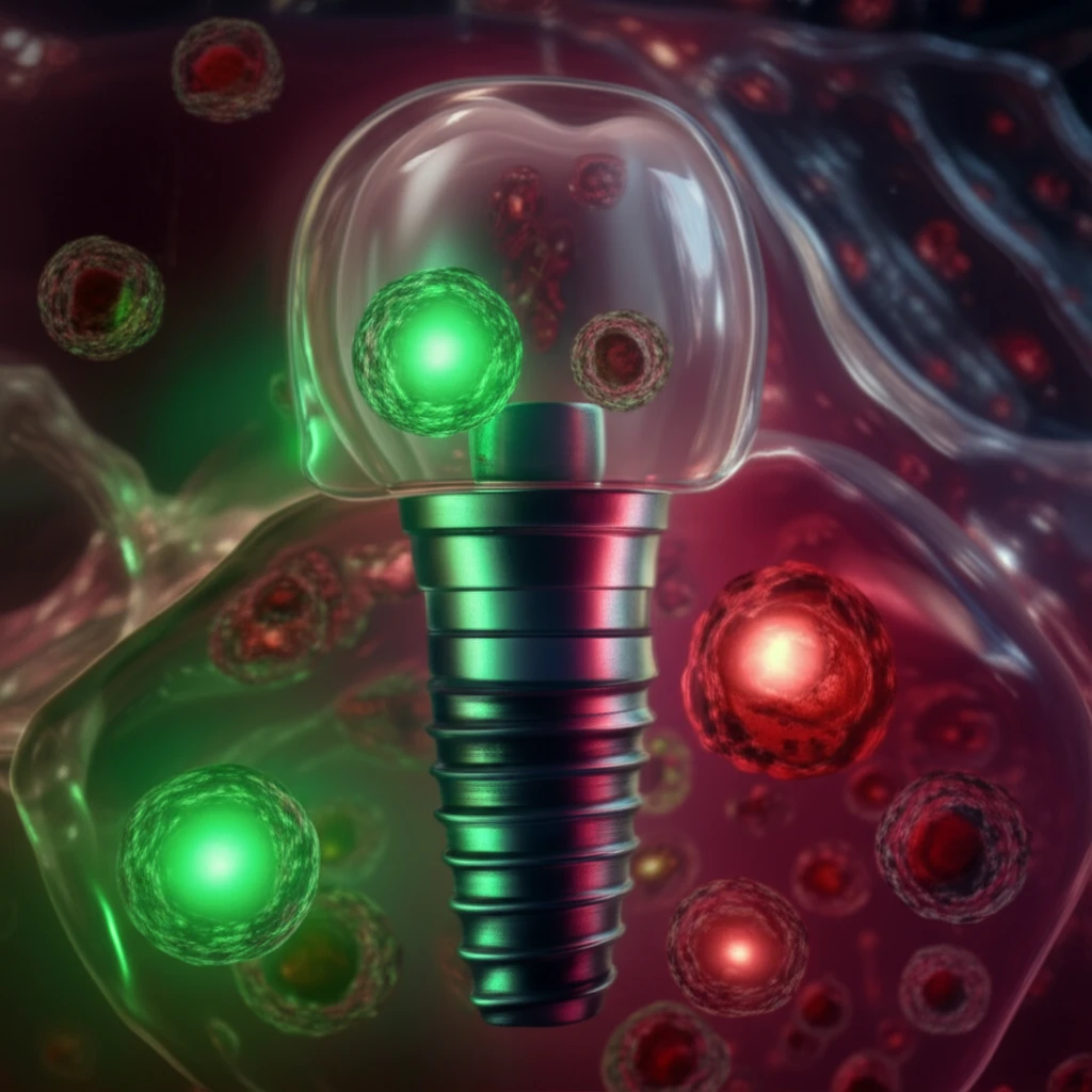
The key innovation lies in the ability to visualize cell death progression in real-time using a non-toxic LIVE/DEAD staining protocol in combination with Scanning Laser Optical Tomography (SLOT). This allows researchers to differentiate between living and dead gingival fibroblasts and monitor their response to various stimuli, such as toxic substances.
- CYTO-ID Red: Stains all human gingival fibroblasts, indicating the total cell population.
- DRAQ7: Enters cells with compromised membranes, binding to DNA and indicating cell death.
- Chlorhexidine: A toxic substance used to induce cell death and validate the LIVE/DEAD staining protocol.
The Future of Dental Implants: Personalized and Optimized
This novel 3D peri-implant model, combined with time-resolved live-cell imaging, represents a significant advancement in dental implant research. It provides a powerful tool for evaluating the performance of novel materials and surfaces, ultimately leading to improved implant designs and reduced risk of peri-implant inflammation.
By offering a more comprehensive understanding of cell behavior in a realistic 3D environment, this technology paves the way for personalized implant solutions tailored to individual patient needs. Future research could explore the interaction of tissue cells with bacterial biofilms, providing insights into preventing biofilm-related implant failures.
While this 3D LIVE/DEAD monitoring is not applicable for high-throughput analysis, the non-invasive method will help monitor cell response on the whole dental implant and complement the typical 2D in vitro investigations of implant materials. The capacity to non-invasively observe dynamic alterations in cell condition on an implant in response to external stress is a substantial step forward that will promote progress in tissue engineering, implant material research, and oral microbiology.