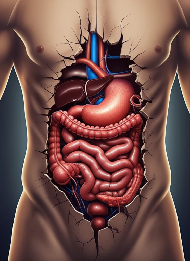
Surreal illustration of thoracoabdominal hernia

When Your Gut Takes a Detour: Understanding Thoracoabdominal Hernias

"A Rare Case Highlights the Importance of Recognizing Unusual Herniations"


Imagine your body as a complex network of interconnected systems, each playing a vital role in maintaining overall health. Now, picture a scenario where one of these systems—your digestive tract—takes an unexpected detour, venturing into a space it doesn't normally occupy. This is essentially what happens in a thoracoabdominal hernia, a rare condition where abdominal organs, like the intestines, push through a defect and migrate into the chest cavity.

Thoracoabdominal hernias are uncommon occurrences, typically arising after significant blunt trauma. In these cases, a rupture in the diaphragm, the muscle separating the chest and abdomen, allows abdominal contents to herniate upwards into the thorax. However, not all hernias follow this conventional pathway. A recent case report details an atypical presentation of this condition, challenging our understanding and highlighting the importance of considering alternative routes of herniation.

This article will delve into a fascinating case of thoracoabdominal herniation that defied expectations. We'll explore the details of the patient's journey, from initial trauma to diagnosis and eventual surgical repair. By examining this unique scenario, we aim to shed light on the complexities of thoracoabdominal hernias and underscore the need for vigilance and tailored treatment approaches.

Atypical Herniation: Bypassing the Diaphragm

Surreal illustration of thoracoabdominal hernia

The case involves a 72-year-old woman with a history of hypertension, hypothyroidism, and asthma who sustained multiple injuries in a road traffic accident. Initial assessments revealed several right-sided issues, including fractures and a haemopneumothorax. Notably, she was also diagnosed with a traumatic hernia involving the hepatic flexure of the colon, protruding through the anterolateral abdominal wall.

Initially, her injuries were managed conservatively, and she was discharged after three weeks. However, upon follow-up, she reported persistent abdominal pain, altered bowel habits, and shortness of breath. Physical examination revealed enlargement of the abdominal wall hernia. Subsequent imaging, specifically computed tomography (CT) colonography, unveiled a surprising progression of the hernia.

The CT scans showed that the hepatic flexure of the colon:
  • Passed through the transversus abdominis and internal oblique muscles.
  • Traveled cranially between the internal and external oblique muscles.
  • Entered the thoracic cavity through a defect in the thoracic wall.
  • Completely bypassed the diaphragm.
This unusual pathway highlights a critical point: thoracoabdominal hernias can manifest in unexpected ways, defying typical anatomical routes. In this case, the bowel didn't pass through the diaphragm, but rather exploited a weakness in the abdominal wall and an intercostal defect to enter the chest cavity. Such atypical presentations necessitate a high degree of clinical suspicion and thorough investigation to ensure accurate diagnosis and appropriate management.

The Importance of Vigilance and Definitive Repair

This unique case underscores the importance of maintaining a high index of suspicion for thoracoabdominal herniation in patients presenting with respiratory or gastrointestinal symptoms following trauma. While imaging plays a crucial role in guiding management, clinicians must be aware of its limitations and the potential for atypical presentations. Early and definitive surgical repair remains the cornerstone of treatment, aiming to restore anatomical integrity and prevent potentially life-threatening complications.

About this Article -

This article was crafted using a human-AI hybrid and collaborative approach. AI assisted our team with initial drafting, research insights, identifying key questions, and image generation. Our human editors guided topic selection, defined the angle, structured the content, ensured factual accuracy and relevance, refined the tone, and conducted thorough editing to deliver helpful, high-quality information.See our About page for more information.

This article is based on research published under:

DOI-LINK: 10.1308/003588414x13814021679032, Alternate LINK

Title: Thoracoabdominal Herniation – But Not As You Know It

Subject: General Medicine

Journal: The Annals of The Royal College of Surgeons of England

Publisher: Royal College of Surgeons of England

Authors: J Wigley, F Noble, A King

Published: 2014-07-01

Everything You Need To Know

1

What exactly is a thoracoabdominal hernia, and what typically causes it?

A thoracoabdominal hernia is a rare condition where abdominal organs, such as the intestines or the hepatic flexure of the colon, protrude through a defect and migrate into the chest cavity. This often occurs after significant blunt trauma that causes a rupture in the diaphragm. However, as demonstrated in the atypical case, herniation can also occur through alternative routes, bypassing the diaphragm entirely and exploiting weaknesses in the abdominal or thoracic wall.

2

How does a typical thoracoabdominal hernia differ from the atypical herniation described, where the diaphragm is bypassed?

In a typical thoracoabdominal hernia, abdominal organs herniate through a rupture in the diaphragm, the muscle separating the chest and abdomen, and enter the thorax. In contrast, the featured case demonstrated an atypical herniation route. The hepatic flexure of the colon bypassed the diaphragm, passing through the transversus abdominis and internal oblique muscles, traveling cranially between the internal and external oblique muscles, and entering the thoracic cavity through a defect in the thoracic wall. This unusual pathway highlights that these hernias can occur in unexpected ways.

3

What are the key steps in diagnosing a thoracoabdominal hernia, especially when an atypical presentation is suspected?

Diagnosis of a thoracoabdominal hernia involves maintaining a high index of suspicion, especially in patients with respiratory or gastrointestinal symptoms following trauma. Imaging techniques like computed tomography (CT) colonography are crucial for visualizing the hernia and determining its extent. However, clinicians must recognize the limitations of imaging and be aware of the potential for atypical presentations where the hernia doesn't follow the typical diaphragmatic route. Thorough investigation is necessary to ensure accurate diagnosis.

4

What is the standard treatment for thoracoabdominal hernias, and why is it considered the cornerstone of care?

Early and definitive surgical repair is the primary treatment for thoracoabdominal hernias. The goal of surgical intervention is to restore anatomical integrity and prevent potentially life-threatening complications. The specific surgical approach may need to be tailored based on the unique characteristics of the hernia, particularly in cases where the herniation route is atypical. The repair addresses the defect in the abdominal or thoracic wall, ensuring that the abdominal organs are properly repositioned and supported.

5

What are the broader implications of atypical thoracoabdominal herniation cases on how clinicians approach diagnosis and treatment?

The atypical case of thoracoabdominal herniation has implications for clinical practice, emphasizing the importance of vigilance and awareness of unusual presentations. Clinicians should consider thoracoabdominal hernia in the differential diagnosis of patients with post-traumatic respiratory or gastrointestinal symptoms, even if there's no obvious diaphragmatic rupture. The case highlights the need for thorough imaging and a tailored surgical approach based on the specific anatomical route of herniation. This understanding can improve diagnostic accuracy and optimize patient outcomes in these rare and complex cases.

Newsletter Subscribe

Subscribe to get the latest articles and insights directly in your inbox.