Weight-Bearing Woes? Unpacking How Foot Radiographs Change Under Pressure
"From subtle shifts to significant changes, discover the impact of weight on your foot's inner workings."
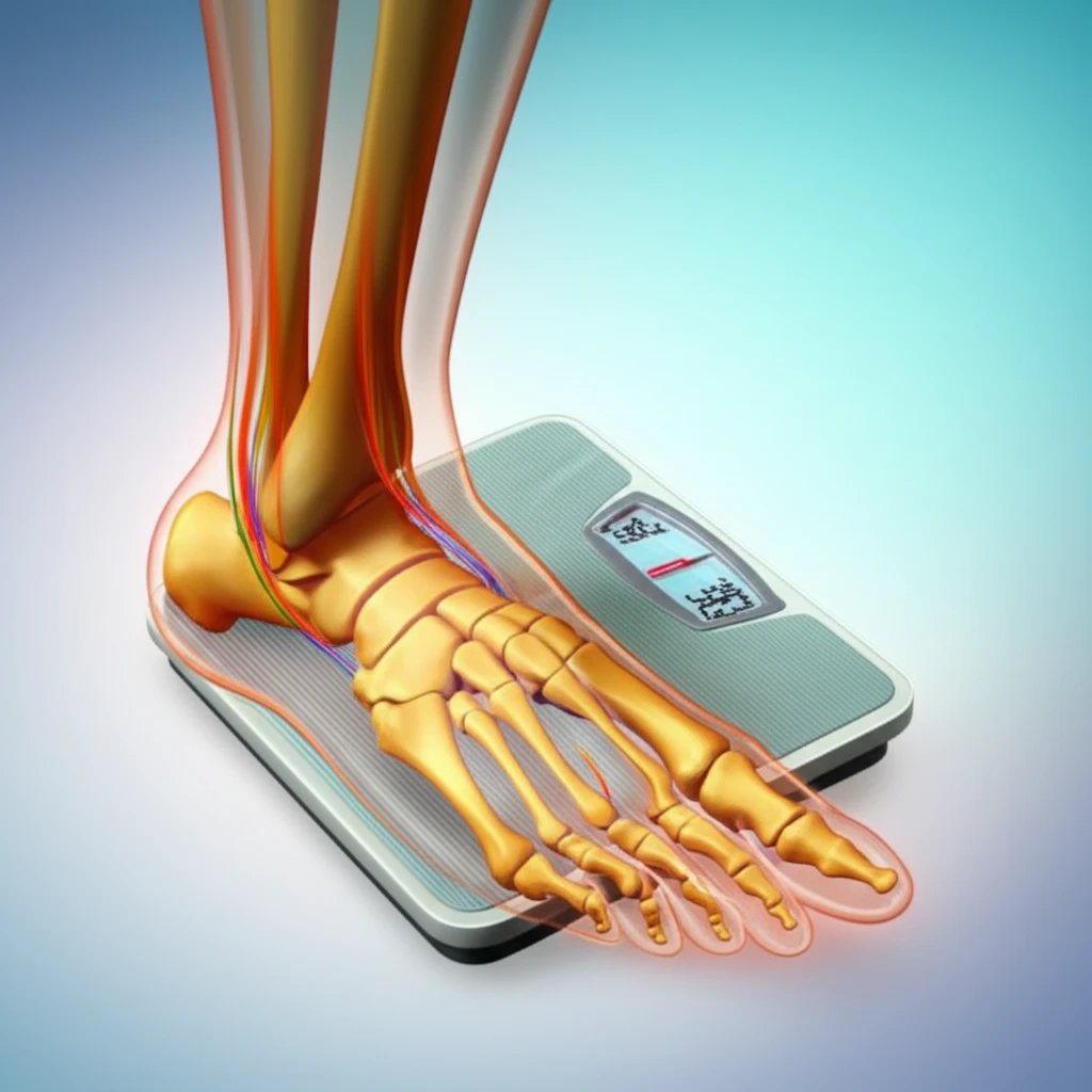
Our feet are marvels of engineering, designed to support our weight and absorb the shocks of movement. But what happens to these intricate structures when we put them under pressure? Clinical decisions about foot health often rely on weight-bearing radiographs, but the impact of varying weight levels on these images has been a subject of scientific inquiry. Recent research has begun to unravel the subtle, yet significant, changes that occur within our feet as we stand, walk, and bear weight.
This article explores a fascinating study that examines how the angle of our big toe (hallux valgus), the space between our foot bones (intermetatarsal angle), and other key measurements shift as we transition from non-weight-bearing to full weight-bearing. By understanding these changes, we can gain valuable insights into foot mechanics and how they relate to common foot conditions and treatments.
Whether you're a healthcare professional, a fitness enthusiast, or simply curious about your body, join us as we delve into the intriguing world of foot radiographs and discover how the simple act of standing can reshape the architecture of your feet.
The Weighty Truth: How Weight-Bearing Changes Foot Radiographs
The study, published in the journal Foot & Ankle Specialist, investigated the effects of different weight-bearing conditions on the feet. Researchers took radiographs of healthy individuals under five distinct conditions: non-weight-bearing, 10% body weight, 25% body weight, 50% body weight, and 100% body weight. They meticulously measured various angles and distances in the foot to see how they changed with each weight level.
- The TNCA and TCA angles increased significantly with increased weight-bearing, indicating arch flattening.
- The CHG decreased with increasing weight, reflecting the arch's descent.
- The HVA and IMA did not change significantly across the different weight-bearing conditions.
Stepping Forward: Implications and Future Directions
This study offers valuable insights into the biomechanics of the foot and how they are influenced by weight. The findings underscore the importance of considering weight-bearing conditions when interpreting foot radiographs. For instance, post-injury or post-surgical rehabilitation protocols might need to consider the level of weight allowed on the foot to assess its accurate structure. Further research is needed to investigate the specific impact of weight-bearing on various foot pathologies. It is important to find how these findings may translate to real-world clinical scenarios to improve diagnoses and optimize patient care. By gaining a deeper understanding of the foot's response to weight, we can take better care of our feet and maintain our mobility for years to come.