Illustration of a heart with a ventricular septal defect and tricuspid regurgitation, showing the defect closing and blood flow normalizing after an intervention.

VSD Closure: How Doctors Fix Tricuspid Regurgitation

"A new ultrasound study reveals how closing ventricular septal defects can improve heart valve function and overall heart health."


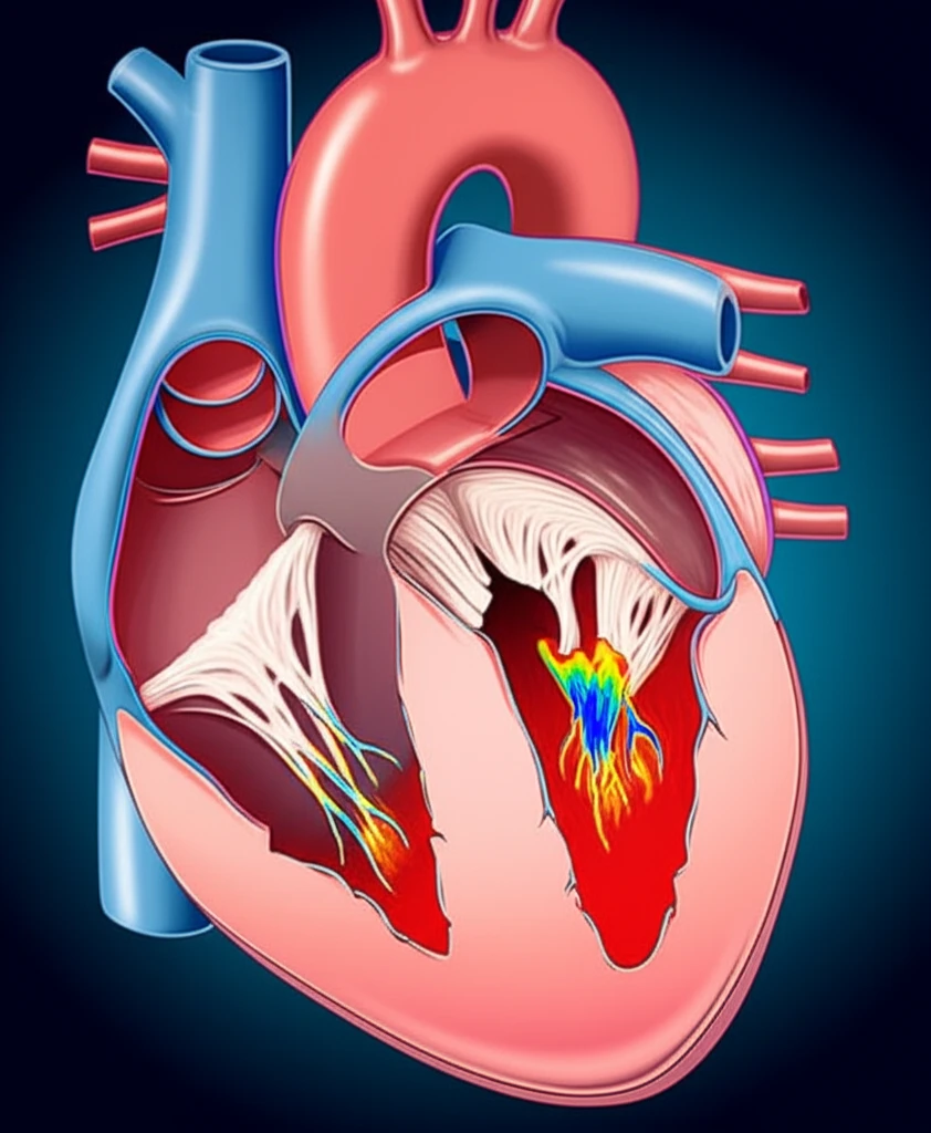
A ventricular septal defect (VSD) is a hole in the wall separating the two lower chambers of the heart. This common congenital heart defect can sometimes lead to other heart problems, including tricuspid regurgitation. Tricuspid regurgitation occurs when the tricuspid valve, which sits between the right atrium and right ventricle, doesn't close properly, causing blood to leak backward.

For simple VSDs, doctors often use a procedure called interventional occlusion to close the hole. While this is generally considered safe, there can be complications, especially when tricuspid regurgitation is also present. It is critical to determine if surgery is the best option.

A new study has explored the connection between VSDs, tricuspid regurgitation, and the effectiveness of VSD occlusion. Using detailed ultrasound imaging, researchers have uncovered key insights into how closing the VSD can improve tricuspid valve function, offering a promising approach to treatment.

Unraveling the Link: VSDs and Tricuspid Regurgitation

Illustration of a heart with a ventricular septal defect and tricuspid regurgitation, showing the defect closing and blood flow normalizing after an intervention.

Researchers used echocardiography (ultrasound of the heart) to study 43 patients with both a membranous VSD and tricuspid regurgitation. They carefully measured the size of the VSD and the amount of tricuspid regurgitation before and after VSD occlusion.

The study revealed four main ways that a VSD can lead to tricuspid regurgitation:

  • Short Tricuspid Valve Septa: The VSD opens directly near the valve, causing blood to flow into the right atrium.
  • Interminable Anterior Tricuspid Valve or Abnormal Chordae Tendineae Attachment: The shunt flow hits the valve or its supporting structures, deflecting blood into the right atrium.
  • Irregular Adhesion of the Right Ventricular Septal Defect: Tissue adhesions create a tunnel, directing blood flow toward the tricuspid valve.
  • Pulmonary Hypertension: Increased pressure in the lungs strains the heart and contributes to tricuspid regurgitation.
Following VSD occlusion, the researchers observed a significant reduction in tricuspid regurgitation volume in most patients. This suggests that closing the VSD can indeed improve the function of the tricuspid valve.

A Promising Future for Heart Health

This research highlights the importance of echocardiography in understanding and treating VSDs complicated by tricuspid regurgitation. By carefully examining the heart's structure and blood flow, doctors can determine the best course of action for each patient.

The study confirms that VSD occlusion can be an effective way to reduce tricuspid regurgitation and improve overall heart health. This minimally invasive procedure offers a safe and promising option for many patients.

With continued research and advancements in treatment techniques, doctors can provide even better care for individuals with VSDs and tricuspid regurgitation, leading to improved outcomes and a better quality of life.

About this Article -

This article was crafted using a human-AI hybrid and collaborative approach. AI assisted our team with initial drafting, research insights, identifying key questions, and image generation. Our human editors guided topic selection, defined the angle, structured the content, ensured factual accuracy and relevance, refined the tone, and conducted thorough editing to deliver helpful, high-quality information.See our About page for more information.

This article is based on research published under:

DOI-LINK: 10.3892/etm.2012.876, Alternate LINK

Title: Investigation Of Membranous Ventricular Septal Defect Complicated With Tricuspid Regurgitation In Ventricular Septal Defect Occlusion

Subject: Cancer Research

Journal: Experimental and Therapeutic Medicine

Publisher: Spandidos Publications

Authors: Shu-Ping Liu, Li Li, Ke-Chun Yao, Na Wang, Jian-Chang Wang

Published: 2012-12-21

Everything You Need To Know

1

What are ventricular septal defects (VSDs) and tricuspid regurgitation?

A ventricular septal defect, or VSD, is a hole in the wall that separates the two lower chambers of the heart. Tricuspid regurgitation happens when the tricuspid valve, which is positioned between the right atrium and right ventricle, does not close correctly. This causes blood to leak backward. While the text focuses on the link between VSD and tricuspid regurgitation it doesn't explain the impact of the leak on the body.

2

How do doctors fix tricuspid regurgitation when it's related to a VSD?

Doctors often use a procedure called interventional occlusion to close a VSD. The ultrasound study showed closing the VSD improved the function of the tricuspid valve. While interventional occlusion is generally considered safe, complications may arise, particularly in the presence of tricuspid regurgitation, making it crucial to assess whether surgery is the best option.

3

What role did ultrasound play in understanding the link between VSDs and tricuspid regurgitation?

Researchers used echocardiography, which is an ultrasound of the heart, to study patients with both a membranous VSD and tricuspid regurgitation. The research looked at the size of the VSD and the amount of tricuspid regurgitation before and after VSD occlusion. These measurements allowed them to understand how closing the VSD affected tricuspid regurgitation.

4

What are the different ways a VSD can cause tricuspid regurgitation, according to recent research?

The study revealed that a VSD can lead to tricuspid regurgitation in several ways including: short tricuspid valve septa, interminable anterior tricuspid valve or abnormal chordae tendineae attachment, irregular adhesion of the right ventricular septal defect and pulmonary hypertension. Each of these impacts the function of the heart and puts strain on the right atrium and right ventricle.

5

What are the implications of this ultrasound study's findings on future treatment of VSDs and related tricuspid regurgitation?

The research showed that closing a VSD through interventional occlusion can significantly reduce tricuspid regurgitation. This is important because reducing the backflow of blood improves the heart's efficiency and overall health. The study shows that doctors can determine the best course of action for each patient by examining the heart's structure and blood flow using echocardiography. However, the text doesn't explain how long the benefits last after the procedure.

Newsletter Subscribe

Subscribe to get the latest articles and insights directly in your inbox.