Vision Under Attack: Unmasking the Rare Fungal Threat of Bipolaris Keratitis
"Learn how this unusual fungal infection can compromise corneal health and what steps can be taken to protect your sight."
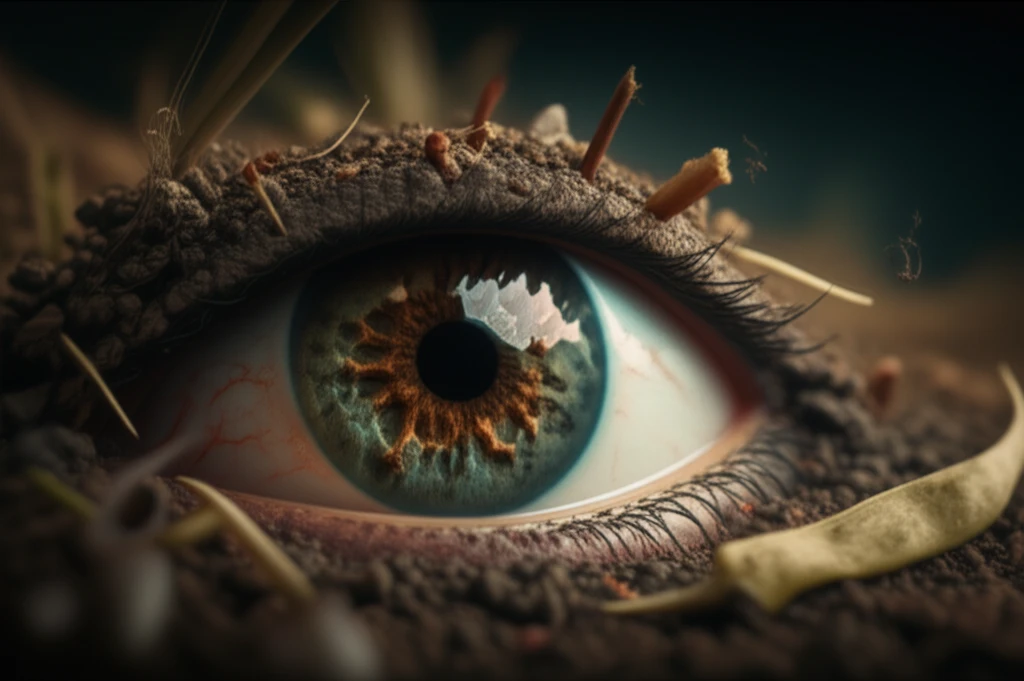
Fungal keratitis represents a significant threat to vision, especially in tropical and subtropical regions, where it is a relatively common cause of corneal infections. The majority of these infections are caused by filamentous fungi, with Aspergillus and Fusarium species being the most commonly identified culprits worldwide. However, a growing concern is the rise of dematiaceous fungi as causative agents of corneal ulcers.
Among these, species of Curvularia and Bipolaris are increasingly recognized as significant contributors to keratomycosis. Bipolaris hawaiiensis, a darkly pigmented fungus, is known to cause subcutaneous, cutaneous, and soft tissue infections referred to as phaeohyphomycosis. While typically associated with plant material, grasses, and soil, Bipolaris hawaiiensis can also lead to bronchopulmonary disease, encephalitis, and, in rare instances, ocular infections.
Given the potential severity of such infections, understanding the causes, symptoms, and treatments for Bipolaris keratitis is crucial for healthcare providers and individuals at risk. Early diagnosis and appropriate management are key to preventing vision loss and improving patient outcomes. This article aims to shed light on the rare but serious threat of Bipolaris keratitis, offering insights into its identification, treatment, and prevention.
What are the Symptoms and How Is It Diagnosed?
A recent case study detailed a 63-year-old male who developed Bipolaris keratitis following a traumatic eye injury involving sawdust. The patient, who already had ocular hypertension in the other eye, initially presented with symptoms that led to a misdiagnosis of a simple corneal ulcer at a local hospital. Despite treatment with amphotericin B, his condition did not improve, necessitating a referral to a specialized ophthalmological department.
- Microscopic Examinations: The corneal lesion was scraped for thorough microscopic analysis, including KOH examination, Gram staining, and calcofluor-white staining, to identify the presence and nature of fungal elements.
- Confocal Microscopy: Confocal microscopy was employed to visualize the structure of the hyphae in real-time, aiding in the identification of fungal characteristics such as acute angle branching.
- Fungal Culture: A fungal culture was performed to grow and identify the specific species of fungi involved, which is critical for tailoring the antifungal treatment.
Protecting Your Sight: The Importance of Early Action
Bipolaris hawaiiensis is a virulent organism that can cause ocular morbidity and blindness, early diagnosis and accurate identification of the pathogenic B. hawaiiensis can lead to prompt and appropriate treatment and prevent ocular morbidity and blindness. In refractory B. hawaiiensis keratomycosis, therapeutic penetrating keratoplasty should be considered to remove the infected corneal tissue and recalcitrant corneal plaque and to improve visual acuity.