Visual representation of interocular suppression, showing competing visual inputs and their impact on the brain.

Vision Mismatch: How Our Eyes Compete and What It Means for Clarity

"New research unveils how the brain juggles conflicting signals from our eyes, impacting vision quality and depth perception, especially in those with binocular vision abnormalities."


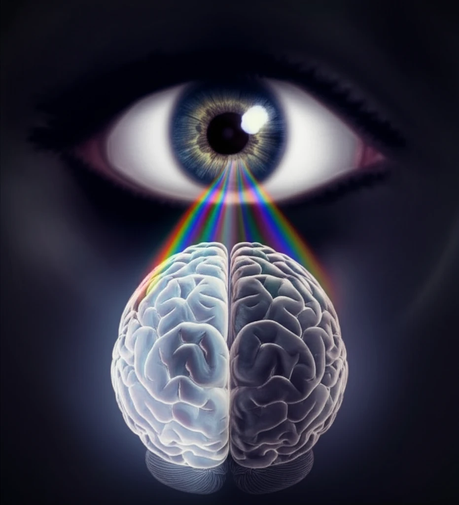
In the realm of sight, what happens when our eyes don't quite see eye-to-eye? Normally, both eyes work in harmony, sending signals to the brain, which then fuses these into a single, clear image. However, this process can be disrupted, leading to a phenomenon called interocular suppression. In essence, when the images seen by each eye differ significantly, the brain suppresses the image from one eye to prevent double vision or confusion. This suppression isn't just a quirk of vision; it's a critical adaptation that influences our depth perception and overall visual clarity.

New research investigates this phenomenon, focusing on individuals with binocular vision abnormalities such as strabismus (crossed eyes) and microstrabismus (a milder form of misalignment). By examining how the brain handles conflicting visual inputs in these conditions, scientists are gaining valuable insights into the mechanisms of interocular suppression and its impact on visual function.

This exploration isn't merely academic; understanding interocular suppression has practical implications for diagnosing and managing vision disorders. For instance, developing more sensitive diagnostic tools could lead to earlier intervention and more effective treatments. Moreover, tailoring visual rehabilitation strategies to address suppression patterns might improve visual outcomes for individuals with binocular vision abnormalities.

Decoding Interocular Suppression: What the Study Reveals

Visual representation of interocular suppression, showing competing visual inputs and their impact on the brain.

A recent study published in the Journal of Vision has shed light on the intricate patterns of interocular suppression in individuals with binocular vision abnormalities. The researchers used specialized visual stimuli to measure the depth and extent of suppression across the central visual field. These stimuli included concentric rings that varied in luminance (brightness) and contrast, allowing the scientists to assess how different types of visual information are processed and suppressed.

The study involved participants with strabismus and microstrabismus, each viewing the stimuli dichoptically—meaning that each eye received a slightly different image. By adjusting the image presented to one eye until it perceptually matched the image in the other eye, the researchers could quantify the degree of suppression occurring.

  • Strabismic Participants: Displayed deeper suppression centrally compared to peripherally, with one hemifield of the visual field experiencing greater suppression than the other.
  • Microstrabismic Participants: Showed weaker suppression than their strabismic counterparts, primarily in the central visual field. However, when contrast-modulated stimuli were used, suppression became broader, deeper, and more pronounced in one hemifield.
  • Stimulus Type Matters: Suppression depth varied depending on the stimulus. Luminance-defined stimuli (L) resulted in deeper suppression than luminance-modulated noise (LM), while contrast-modulated noise (CM) led to deeper suppression than LM stimuli.
Furthermore, the study found a positive correlation between the depth of suppression and both interocular visual acuity difference and stereoacuity reduction. This suggests that greater disparities between the eyes' visual input lead to stronger suppression and poorer depth perception.

Looking Ahead: Clinical Applications and Future Research

These findings have significant implications for clinical practice. The study suggests that LM stimuli could be valuable for assessing suppression patterns in individuals with deep amblyopia (lazy eye), while CM stimuli might be more sensitive for detecting milder suppression in those with microstrabismus. Ultimately, a better understanding of interocular suppression can lead to more targeted and effective vision therapy, helping individuals with binocular vision abnormalities achieve clearer, more comfortable vision.

About this Article -

This article was crafted using a human-AI hybrid and collaborative approach. AI assisted our team with initial drafting, research insights, identifying key questions, and image generation. Our human editors guided topic selection, defined the angle, structured the content, ensured factual accuracy and relevance, refined the tone, and conducted thorough editing to deliver helpful, high-quality information.See our About page for more information.

Everything You Need To Know

1

What is interocular suppression and why is it important?

Interocular suppression is the brain's mechanism for dealing with conflicting visual signals from each eye. When the images seen by each eye are too different, the brain suppresses the image from one eye to prevent double vision or visual confusion. This process is critical for maintaining clear vision and depth perception, particularly in individuals with binocular vision abnormalities.

2

How do binocular vision abnormalities like strabismus and microstrabismus affect interocular suppression?

Binocular vision abnormalities, such as strabismus (crossed eyes) and microstrabismus (a milder form of misalignment), can disrupt the normal binocular vision. In these conditions, the eyes do not work together to create a single, clear image. The brain relies more heavily on interocular suppression to manage the conflicting visual inputs from each eye. This can lead to a reduced visual acuity in one eye and impaired depth perception.

3

How was interocular suppression measured in the recent study?

The recent study used specialized visual stimuli, including concentric rings that varied in luminance (brightness) and contrast, to measure the depth and extent of interocular suppression across the visual field. Participants with strabismus and microstrabismus viewed these stimuli dichoptically, meaning each eye received a slightly different image. By adjusting the image presented to one eye until it matched the image in the other, the researchers quantified the degree of suppression occurring.

4

What were the key findings of the study regarding interocular suppression in participants with strabismus and microstrabismus?

The study found that individuals with strabismus displayed deeper suppression centrally compared to peripherally, with one side of the visual field experiencing greater suppression than the other. Those with microstrabismus showed weaker suppression, primarily in the central visual field. The study also found that suppression depth varied depending on the type of stimulus used. These findings highlight the complex nature of interocular suppression and its relationship to binocular vision abnormalities.

5

What are the clinical implications of the study's findings on interocular suppression?

The research suggests that different types of visual stimuli could be valuable for assessing suppression patterns in those with binocular vision abnormalities. For example, luminance-modulated (LM) stimuli might be useful for assessing suppression in individuals with deep amblyopia (lazy eye), while contrast-modulated (CM) stimuli may be more sensitive for detecting milder suppression in those with microstrabismus. This understanding can lead to more targeted and effective vision therapy.

Newsletter Subscribe

Subscribe to get the latest articles and insights directly in your inbox.