Microfluidic device with glowing endothelial cells

Vein-on-a-Chip: The Future of Cardiovascular Research is Here

"Researchers develop a simple microfluidic device that mimics a blood vessel, offering a cost-effective and efficient platform for cell culture, drug testing, and vascular studies."


In multicellular organisms, cells are organized three-dimensionally (3D) into cooperative assemblies, forming tissues and organs. Within this microenvironment, cells experience dynamic variations like chemical gradients and mechanical forces, which are crucial for their growth, survival, and function. Understanding how cells and tissues interact is critical for investigating human pathology.

Traditional cell cultures are essential in cell biology, biochemistry, and drug discovery. However, two-dimensional (2D) monolayer cell cultures and animal models often fail to accurately replicate the complex structure, function, and physiology of living tissues. This limitation has spurred the development of new in vitro models that better mimic organ functionality, with microsystems engineering playing a crucial role.

Microfabrication techniques enable the creation of microchips that allow precise control over cell position, function, and tissue organization. When combined with microfluidic technology, these microchips provide dynamic control over fluid flow and pressure, creating a microenvironment that closely mimics physiological conditions. This has led to the development of organs-on-chips, which are devices that allow the study of biological processes in vitro that were previously impossible to observe.

What is the Vein-on-a-Chip and How Does It Work?

Microfluidic device with glowing endothelial cells

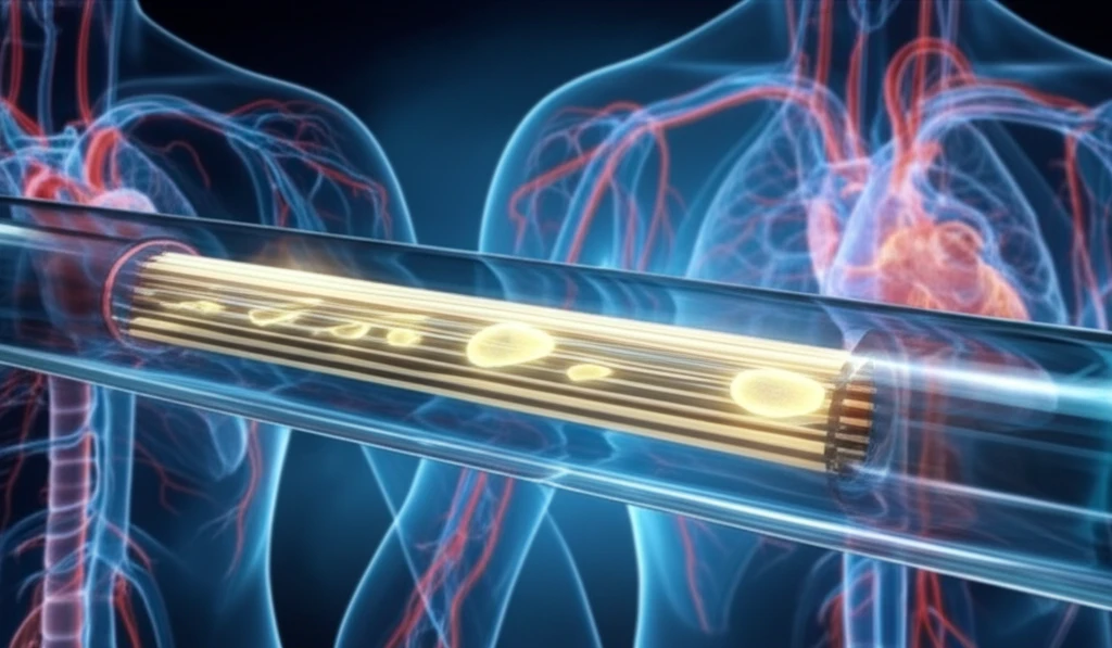
Researchers have successfully created a microfluidic device that mimics a blood vessel using an inexpensive and straightforward method. This "vein-on-a-chip" serves as a starting point for cell culture under perfusion, cardiovascular research, and toxicological studies.

The device is made of polyester and toner (PT), which were tested for cytotoxicity. Endpoint assays revealed that these materials do not induce cell death or nitric oxide (NO) production. To improve endothelial cell adhesion and proliferation, the microchannel was treated with oxygen plasma and fibronectin.

  • Polyester-Toner Microfluidic Device: An inexpensive and easy-to-produce microfluidic device that mimics a blood vessel.
  • Material Safety: Made of polyester and toner (PT), confirmed not to induce cell death or nitric oxide (NO) production.
  • Surface Treatment: Applying oxygen plasma and fibronectin improves endothelial cell adhesion and proliferation along the microchannel.
  • VEGF-A Increase: Treatments increase vascular endothelial growth factor (VEGF-A) concentration profiles, promoting endothelialization for neovascularization.
The simplicity of the device makes it a powerful tool for those who need a rapid microfabrication method in cell biology or organ-on-a-chip research. This "vein-on-a-chip" model has the potential to transform how scientists study blood vessels and develop new treatments for cardiovascular diseases and other related conditions.

The Future of Vein-on-a-Chip Technology

The development of this vein-on-a-chip technology marks a significant step forward in cardiovascular research. Its simplicity, cost-effectiveness, and ability to mimic blood vessel conditions make it an invaluable tool for cell culture, drug testing, and understanding vascular diseases. As research continues, this technology promises to accelerate discoveries and improve treatments for a wide range of conditions.

About this Article -

This article was crafted using a human-AI hybrid and collaborative approach. AI assisted our team with initial drafting, research insights, identifying key questions, and image generation. Our human editors guided topic selection, defined the angle, structured the content, ensured factual accuracy and relevance, refined the tone, and conducted thorough editing to deliver helpful, high-quality information.See our About page for more information.

Everything You Need To Know

1

What is a Vein-on-a-Chip and what are its primary applications?

The Vein-on-a-Chip is a microfluidic device designed to mimic a blood vessel. It is constructed using polyester and toner (PT) through a simple and cost-effective method. Its primary applications include cell culture, drug testing, and vascular studies. It offers a platform to study endothelial cell behavior, assess drug effects, and investigate vascular diseases in a controlled microenvironment that closely resembles the conditions within a blood vessel. This allows researchers to conduct experiments that provide more accurate and relevant results compared to traditional 2D cell cultures or animal models.

2

How does the Vein-on-a-Chip improve upon traditional methods in cardiovascular research?

Traditional methods, such as 2D cell cultures and animal models, often fail to accurately replicate the complex structure, function, and physiology of living tissues. The Vein-on-a-Chip overcomes these limitations by creating a 3D microenvironment that mimics the dynamic conditions of a blood vessel. It allows for precise control over cell position, function, and tissue organization. The use of microfluidic technology provides dynamic control over fluid flow and pressure, closely mimicking physiological conditions. This innovative approach allows for more accurate and relevant results compared to previous methods.

3

What materials are used in the construction of the Vein-on-a-Chip and what safety measures are taken?

The Vein-on-a-Chip is made of polyester and toner (PT). The safety of these materials is ensured through cytotoxicity testing. Endpoint assays were performed to confirm that these materials do not induce cell death or nitric oxide (NO) production, ensuring that the device is biocompatible and will not harm the cells being studied. Furthermore, the microchannel is treated with oxygen plasma and fibronectin to improve endothelial cell adhesion and proliferation, enhancing the device's ability to support cell growth and function.

4

How is the Vein-on-a-Chip optimized to support endothelial cell growth and function?

To optimize the Vein-on-a-Chip for endothelial cell growth and function, the microchannel is treated with oxygen plasma and fibronectin. These treatments enhance the adhesion and proliferation of endothelial cells within the device. Additionally, treatments increase vascular endothelial growth factor (VEGF-A) concentration profiles, promoting endothelialization and neovascularization. This ensures that the cells thrive within the device, allowing for more accurate and meaningful research into blood vessel behavior and disease.

5

What is the significance of VEGF-A in the context of the Vein-on-a-Chip, and what are the long-term implications of this technology?

VEGF-A (vascular endothelial growth factor A) plays a critical role in the Vein-on-a-Chip because it promotes endothelialization and neovascularization. The treatments applied to the device increase VEGF-A concentration profiles, encouraging the formation of new blood vessels. Long-term, the development of the Vein-on-a-Chip technology marks a significant step forward in cardiovascular research. Its simplicity, cost-effectiveness, and ability to mimic blood vessel conditions make it an invaluable tool for cell culture, drug testing, and understanding vascular diseases. This technology promises to accelerate discoveries and improve treatments for a wide range of conditions related to blood vessels.

Newsletter Subscribe

Subscribe to get the latest articles and insights directly in your inbox.