Urologic Imaging: Advances and Expert Insights for Better Health
"Explore the cutting-edge world of urologic imaging, understand how it's improving diagnostics, and learn about the specialists who are leading the way."
Urologic imaging has undergone a remarkable transformation, evolving into a sophisticated field that plays a pivotal role in modern healthcare. From detecting subtle anomalies to guiding complex surgical procedures, advanced imaging techniques provide invaluable insights into the urinary system and related structures. This article explores the key advances in urologic imaging, highlighting the experts and technologies that are improving patient outcomes.
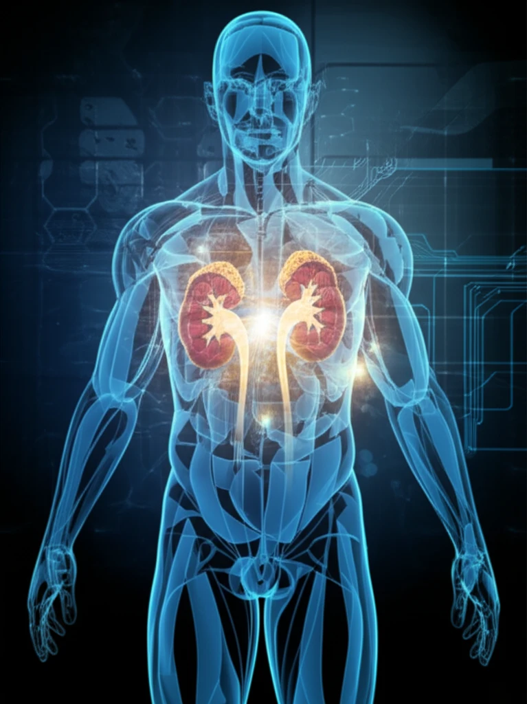
The ability to visualize the intricate details of the kidneys, bladder, prostate, and other urologic organs has revolutionized diagnostics. Techniques like MRI, CT scans, ultrasound, and nuclear medicine imaging allow clinicians to identify tumors, stones, infections, and other abnormalities with greater precision than ever before. Early and accurate detection is often the key to successful treatment and improved quality of life.
This comprehensive overview draws upon the expertise of leading radiologists, urologists, and oncologists who are at the forefront of urologic imaging. Their collective knowledge and experience offer a unique perspective on the current state of the field and the exciting developments on the horizon. Discover how these specialists are collaborating to push the boundaries of what's possible, ultimately benefiting patients around the world.
The Experts Behind the Image: Key Contributors in Urologic Imaging
The field of urologic imaging is driven by a diverse group of specialists, each contributing their unique skills and knowledge to advance the field. These experts, including radiologists, urologists, oncologists, and nuclear medicine physicians, work collaboratively to ensure accurate diagnoses and effective treatment plans.
- Radiologists: Interpreting images, performing minimally invasive procedures, and guiding biopsies.
- Urologists: Using imaging to diagnose and manage urologic conditions, perform surgery, and monitor treatment progress.
- Oncologists: Utilizing imaging to stage and monitor urologic cancers, assess treatment response, and detect recurrence.
- Nuclear Medicine Physicians: Employing molecular imaging techniques to visualize metabolic processes and identify disease at the cellular level.
The Future of Urologic Imaging: Promising Directions
As technology continues to advance, urologic imaging is poised to become even more sophisticated and precise. Emerging techniques such as artificial intelligence (AI) and machine learning (ML) are being integrated into imaging workflows to improve image analysis, enhance detection rates, and personalize treatment strategies.
AI algorithms can be trained to identify subtle patterns and anomalies that may be missed by the human eye, leading to earlier and more accurate diagnoses. ML models can also predict treatment response and tailor therapies to individual patient characteristics, optimizing outcomes and minimizing side effects.
The ongoing collaboration between researchers, clinicians, and industry partners is fueling innovation and driving the field forward. With a continued focus on improving image quality, reducing radiation exposure, and expanding the applications of molecular imaging, urologic imaging promises to play an increasingly important role in the management of urologic diseases in the years to come.