Foot transforming into an X-ray under increasing weight.

Unveiling the Truth: How Weight-Bearing Impacts Your Foot Radiographs

"New research sheds light on how different levels of weight-bearing affect foot X-rays, potentially changing how we diagnose and treat foot conditions."


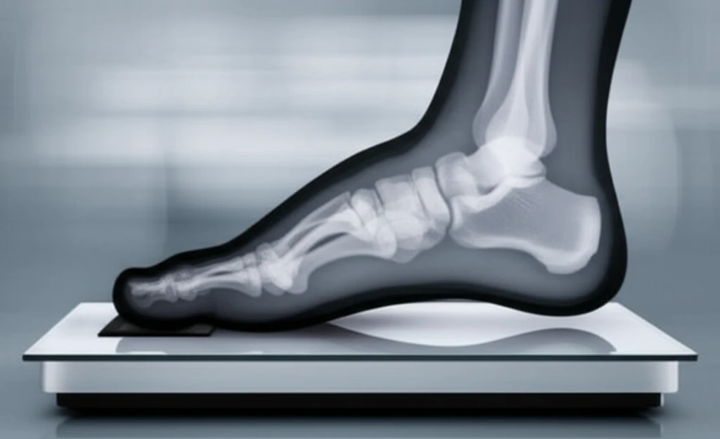
Foot radiographs, or X-rays, are a cornerstone of diagnosing and managing various foot conditions. Often, clinical decisions hinge on these weight-bearing images. But have you ever wondered how the amount of weight placed on the foot during the X-ray affects the results? A recent study delves into this very question, revealing surprising insights into how different weight-bearing conditions can alter radiographic measurements.

The study, published in Foot & Ankle Specialist, sought to determine whether varying percentages of weight-bearing influence radiographic measurements of the normal foot. By examining a group of healthy individuals under five different weight-bearing conditions, researchers uncovered significant changes in specific measurements, challenging some common assumptions about foot imaging.

For anyone who's experienced foot pain, is recovering from an injury, or is simply curious about foot health, these findings could have a significant impact. Let's explore the key takeaways from this research and what they mean for understanding your foot radiographs.

Decoding Weight-Bearing: What the Study Revealed

Foot transforming into an X-ray under increasing weight.

The prospective study involved 20 healthy participants who underwent foot radiographs under five conditions: non-weight-bearing, 10% body weight, 25% body weight, 50% body weight, and 100% body weight. Researchers then meticulously measured several key radiographic parameters, including:

These measurements aimed to capture a comprehensive picture of foot alignment and structure under varying degrees of stress.

  • Hallux valgus angle (HVA)
  • 1-2 intermetatarsal angle (IMA)
  • Talonavicular coverage angle (TNCA)
  • Talocalcaneal angle (TCA)
  • Forefoot width
  • LisFranc distance
  • Cuboid height to ground (CHG)
  • Talo-first metatarsal angle (TMA)
The results revealed some noteworthy shifts:
  • TNCA and TCA Increased: These angles, indicative of midfoot abduction and hindfoot alignment, respectively, significantly increased as weight-bearing increased.
  • CHG Decreased: The cuboid height to ground, a measure of arch height, decreased with increasing weight-bearing, suggesting a flattening of the medial arch.
  • No Change in HVA, IMA, Forefoot Width, LisFranc Distance, and TMA: These measurements remained relatively stable regardless of the weight-bearing condition.

The Bottom Line: What This Means for You

This research highlights the importance of considering weight-bearing conditions when interpreting foot radiographs. The study suggests that radiographic changes in the foot occur primarily between non-weight-bearing and 25% weight-bearing. Once a patient is bearing at least 25% of their body weight on their foot, the radiographs would be no different than if they were standing on one leg.

About this Article -

This article was crafted using a human-AI hybrid and collaborative approach. AI assisted our team with initial drafting, research insights, identifying key questions, and image generation. Our human editors guided topic selection, defined the angle, structured the content, ensured factual accuracy and relevance, refined the tone, and conducted thorough editing to deliver helpful, high-quality information.See our About page for more information.

Everything You Need To Know

1

What is the primary focus of the study regarding foot radiographs?

The study primarily focused on determining how different levels of weight-bearing affect the radiographic measurements of the foot. It investigated the changes in foot X-rays under various weight-bearing conditions, such as non-weight-bearing, 10%, 25%, 50%, and 100% body weight, to understand their impact on diagnostic accuracy and treatment decisions.

2

What specific radiographic parameters were measured in the study, and what do they represent?

The study measured several key radiographic parameters to assess foot alignment and structure under varying degrees of stress. These included the Hallux valgus angle (HVA), 1-2 intermetatarsal angle (IMA), Talonavicular coverage angle (TNCA), Talocalcaneal angle (TCA), Forefoot width, LisFranc distance, Cuboid height to ground (CHG), and Talo-first metatarsal angle (TMA). Each parameter provides specific information: HVA and IMA relate to the alignment of the big toe and metatarsals, TNCA and TCA reflect midfoot and hindfoot alignment, CHG indicates arch height, and the others offer additional insights into foot structure.

3

How did weight-bearing influence the measurements of TNCA, TCA, and CHG in the study?

The study found that TNCA and TCA, which indicate midfoot abduction and hindfoot alignment, significantly increased as weight-bearing increased. This suggests changes in foot structure under load. Conversely, CHG, a measure of arch height, decreased with increasing weight-bearing, indicating flattening of the medial arch under load.

4

What are the implications of this study's findings for interpreting foot radiographs in clinical practice?

The research underscores the importance of considering weight-bearing conditions when interpreting foot radiographs. The study suggests that significant changes in radiographic measurements occur primarily between non-weight-bearing and 25% weight-bearing. The study implies that once a patient is bearing at least 25% of their body weight on their foot, the radiographs would be no different than if they were standing on one leg. This could influence how healthcare professionals diagnose conditions, monitor the progression of foot problems, and make decisions about treatment.

5

Why is it important to understand how weight-bearing affects foot radiographs, and how can this knowledge improve patient care?

Understanding the influence of weight-bearing on foot radiographs is crucial for accurate diagnosis and effective treatment of foot conditions. As the study revealed, different weight-bearing conditions can alter radiographic measurements. By considering these variations, clinicians can avoid misinterpretations and make more informed decisions. This can lead to more precise diagnoses, personalized treatment plans, and improved patient outcomes. Awareness of weight-bearing's impact ensures that the foot radiographs provide a more complete and accurate picture of the patient's foot health, aiding in better care and management of various foot-related issues.

Newsletter Subscribe

Subscribe to get the latest articles and insights directly in your inbox.