3D representation of collagen fiber orientation within a carotid plaque

Unveiling the Hidden Architecture of Carotid Plaques: What 3D Fiber Orientation Means for Your Health

"New research sheds light on the intricate collagen structure within carotid plaques, potentially revolutionizing our understanding and treatment of stroke risk."


Stroke remains a leading cause of disability and death worldwide, and a major culprit behind many strokes is the rupture of atherosclerotic plaques in the carotid arteries. These plaques, built up over time from cholesterol, calcium, and other substances, can become unstable and break off, leading to blockages in the brain. Understanding what makes a plaque vulnerable to rupture is crucial for developing effective prevention strategies.

For years, researchers have been investigating the composition and structure of these plaques, focusing on factors like size, lipid content, and the presence of inflammatory cells. One key component that has garnered increasing attention is collagen, a fibrous protein that provides strength and stability to tissues throughout the body. In carotid plaques, the arrangement and orientation of collagen fibers are thought to play a critical role in determining whether a plaque will remain stable or become prone to rupture.

Now, a new study published in the Journal of Structural Biology is offering unprecedented insights into the 3D architecture of collagen fibers within carotid plaques. Using a sophisticated imaging technique called Diffusion Tensor Imaging (DTI), researchers have mapped the orientation of these fibers in detail, revealing patterns that could help identify high-risk plaques and pave the way for more targeted treatments.

The Collagen Connection: Why Fiber Orientation Matters

3D representation of collagen fiber orientation within a carotid plaque

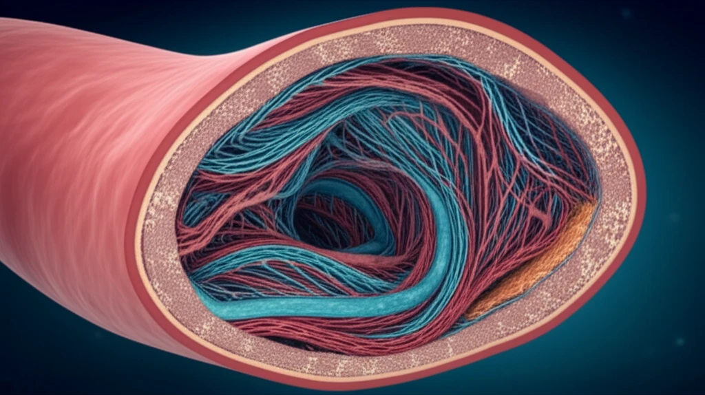
Collagen acts as the main structural protein in blood vessels, similar to steel beams in a building. These fibers are oriented in specific ways to withstand the forces exerted on the artery walls. In healthy arteries, collagen fibers typically align circumferentially, providing resistance to the pressure of blood flow. However, in atherosclerotic plaques, this orderly arrangement can become disrupted.

Previous studies have primarily relied on 2D histological examinations, providing a limited view of collagen fiber orientation. This new research, utilizing DTI, offers a comprehensive 3D perspective, allowing scientists to analyze the complex interplay of fiber arrangements within the entire plaque structure. The DTI technique works by measuring the diffusion of water molecules within the tissue. Water diffuses more readily along the direction of the fibers, allowing researchers to infer the fiber orientation.

  • Circumferential Orientation: Fibers aligned around the circumference of the artery, providing support against blood pressure.
  • Longitudinal Orientation: Fibers aligned along the length of the artery, potentially offering resistance to stretching and bending.
  • Radial Orientation: Fibers oriented from the inner to outer layers of the plaque.
The study, led by researchers at Erasmus Medical Center and Eindhoven University of Technology, involved imaging seven carotid plaques removed from patients undergoing endarterectomy (surgical removal of plaque). Using a high-powered 9.4 Tesla MRI scanner, the team applied DTI to map the 3D orientation of collagen fibers within the plaques. The results revealed a fascinating picture of plaque architecture.

The Road Ahead: Translating Research into Better Patient Care

This study marks a significant step forward in our understanding of carotid plaque biology. By providing a detailed map of 3D collagen fiber orientation, the research opens up new avenues for assessing plaque vulnerability and developing more effective prevention strategies. Further research is needed to validate these findings in larger patient cohorts and to explore the potential for using DTI as a non-invasive tool for identifying high-risk plaques in vivo. Ultimately, the goal is to translate this knowledge into better patient care, reducing the burden of stroke and improving cardiovascular health for all.

About this Article -

This article was crafted using a human-AI hybrid and collaborative approach. AI assisted our team with initial drafting, research insights, identifying key questions, and image generation. Our human editors guided topic selection, defined the angle, structured the content, ensured factual accuracy and relevance, refined the tone, and conducted thorough editing to deliver helpful, high-quality information.See our About page for more information.

This article is based on research published under:

DOI-LINK: 10.1016/j.jsb.2017.08.003, Alternate LINK

Title: 3D Fiber Orientation In Atherosclerotic Carotid Plaques

Subject: Structural Biology

Journal: Journal of Structural Biology

Publisher: Elsevier BV

Authors: Ali C. Akyildiz, Chen-Ket Chai, Cees W.J. Oomens, Aad Van Der Lugt, Frank P.T. Baaijens, Gustav J. Strijkers, Frank J.H. Gijsen

Published: 2017-10-01

Everything You Need To Know

1

What are carotid plaques, and why are they important?

Atherosclerotic plaques in the carotid arteries are built up over time from cholesterol, calcium, and other substances. These plaques can become unstable and rupture, leading to blockages in the brain and causing strokes. The composition and structure of these plaques, including factors like size, lipid content, and the presence of inflammatory cells, are all critical in understanding how they contribute to stroke risk.

2

What is the role of collagen in carotid plaques?

Collagen is a fibrous protein that provides strength and stability to tissues, including the carotid arteries. In the context of carotid plaques, the arrangement and orientation of collagen fibers play a crucial role in determining plaque stability. The alignment of collagen fibers is thought to influence whether a plaque will remain stable or become prone to rupture, which can lead to stroke. Disruptions in the typical fiber arrangements can weaken the plaque.

3

How does Diffusion Tensor Imaging (DTI) work in this context?

Diffusion Tensor Imaging (DTI) is a sophisticated imaging technique used to map the 3D orientation of collagen fibers within carotid plaques. DTI works by measuring the diffusion of water molecules within the tissue. Water diffuses more readily along the direction of the fibers, allowing researchers to infer the fiber orientation. This technique provides a comprehensive 3D perspective of the collagen fiber arrangement, unlike previous 2D methods.

4

What are the different orientations of collagen fibers that were identified?

The study identified several key orientations of collagen fibers. Circumferential orientation, where fibers are aligned around the circumference of the artery, provides support against blood pressure. Longitudinal orientation involves fibers aligned along the length of the artery, offering resistance to stretching and bending. Radial orientation refers to fibers oriented from the inner to outer layers of the plaque. These orientations collectively contribute to the overall structural integrity and stability of the plaque.

5

Who conducted the study, and what is the ultimate goal of this research?

The research, conducted by researchers at Erasmus Medical Center and Eindhoven University of Technology, involved imaging seven carotid plaques removed from patients using DTI. Using a high-powered 9.4 Tesla MRI scanner, they mapped the 3D orientation of collagen fibers within the plaques. The aim is to translate this knowledge into better patient care, reducing the burden of stroke and improving cardiovascular health. Further research is needed to validate these findings in larger patient cohorts and to explore the potential for using DTI as a non-invasive tool for identifying high-risk plaques in vivo.

Newsletter Subscribe

Subscribe to get the latest articles and insights directly in your inbox.