3D CE-FLAIR imaging highlights a brain biopsy site within leptomeningeal lesions.

Untangling Neurosarcoidosis: How Advanced Imaging Can Lead to Accurate Diagnosis

"Discover the vital role of contrast-enhanced 3D FLAIR imaging in diagnosing neurosarcoidosis, offering new hope for those with chronic meningitis."


Neurosarcoidosis, a rare inflammatory condition affecting the nervous system, presents a significant diagnostic challenge. It manifests in 5% to 15% of sarcoidosis patients, mimicking conditions like tuberculosis, lymphoma, and vasculitis. Accurate diagnosis hinges on detailed central nervous system histology, which is often invasive.

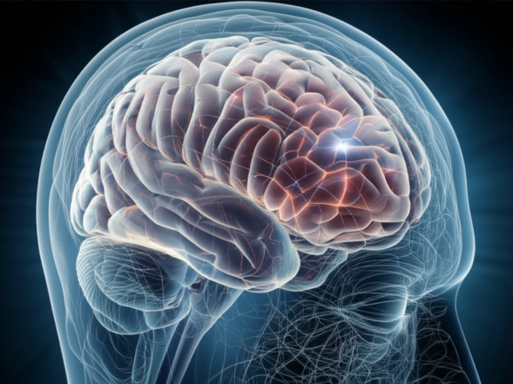
Fluid-attenuated inversion recovery (FLAIR) imaging, especially when contrast-enhanced, has emerged as a valuable tool. By suppressing cerebrospinal fluid signals and highlighting blood-brain barrier disruptions, CE-FLAIR enhances the visibility of leptomeningeal lesions often missed by standard imaging techniques.

This article explores how contrast-enhanced 3D FLAIR imaging can be a game-changer in diagnosing neurosarcoidosis. By presenting a real-world case, we'll demonstrate how this advanced technique aids in selecting optimal brain biopsy sites, leading to definitive diagnoses in cases of chronic meningitis with undetermined causes.

3D CE-FLAIR: A Clearer Picture for Brain Biopsy

3D CE-FLAIR imaging highlights a brain biopsy site within leptomeningeal lesions.

Traditional diagnostic methods for neurosarcoidosis can be inconclusive. While CE-T1-weighted imaging is commonly used, it sometimes fails to detect subtle leptomeningeal lesions. This is where contrast-enhanced 3D FLAIR (CE-FLAIR) imaging steps in, offering a more detailed and sensitive approach.

A recent case highlighted the utility of CE-FLAIR. A 46-year-old woman with chronic headaches, fever, and diplopia underwent extensive testing, including CSF analysis, which revealed pleocytosis and elevated ACE and ADA levels. However, the underlying cause remained elusive.

  • Initial MRI scans with CE-T1-weighted imaging showed some meningeal lesions, but 3D CE-FLAIR imaging provided a much clearer view, particularly in the cerebellar region.
  • The enhanced visualization from 3D CE-FLAIR guided a precise brain biopsy, targeting a specific lesion in the right cerebellum.
  • Histological examination of the biopsied tissue confirmed the presence of non-caseating granulomas, a hallmark of neurosarcoidosis.
This case underscores the superiority of 3D CE-FLAIR in detecting leptomeningeal lesions suitable for biopsy. Its ability to minimize partial-volume effects and suppress CSF flow artifacts allows for better visualization of subtle abnormalities, leading to more accurate diagnoses and timely treatment.

The Future of Neurosarcoidosis Diagnosis

The successful diagnosis in this case highlights the potential of 3D CE-FLAIR imaging to improve diagnostic accuracy and patient outcomes. By providing a clearer view of leptomeningeal lesions, this technique enables clinicians to make informed decisions about biopsy sites, reducing the risk of complications and delays in treatment.

While further research is needed to validate these findings, 3D CE-FLAIR imaging holds promise as a valuable tool in the diagnostic workup of chronic meningitis, particularly when neurosarcoidosis is suspected. Its non-invasive nature and enhanced visualization capabilities make it an attractive alternative to more invasive procedures.

For individuals experiencing unexplained neurological symptoms, including chronic headaches, fever, and visual disturbances, consulting with a neurologist and exploring advanced imaging options like 3D CE-FLAIR may be crucial in obtaining an accurate diagnosis and initiating appropriate treatment.

About this Article -

This article was crafted using a human-AI hybrid and collaborative approach. AI assisted our team with initial drafting, research insights, identifying key questions, and image generation. Our human editors guided topic selection, defined the angle, structured the content, ensured factual accuracy and relevance, refined the tone, and conducted thorough editing to deliver helpful, high-quality information.See our About page for more information.

This article is based on research published under:

DOI-LINK: 10.1016/j.clineuro.2018.08.006, Alternate LINK

Title: Clinical Diagnostic Utility Of Contrast-Enhanced Three-Dimensional Fluid-Attenuated Inversion Recovery For Selection Of Brain Biopsy Sites In Neurosarcoidosis: A Case Report

Subject: Neurology (clinical)

Journal: Clinical Neurology and Neurosurgery

Publisher: Elsevier BV

Authors: Tatsuya Ueno, Rie Desaki, Tomoya Kon, Rie Haga, Jin-Ichi Nunomura, Kensuke Murakami, Masahiko Tomiyama

Published: 2018-10-01

Everything You Need To Know

1

What is neurosarcoidosis?

Neurosarcoidosis is a rare inflammatory condition affecting the nervous system, occurring in 5% to 15% of sarcoidosis patients. This condition is significant because it can mimic other diseases like tuberculosis, lymphoma, and vasculitis, making accurate diagnosis challenging. Precise identification is crucial for initiating timely and appropriate treatment, which can greatly impact patient outcomes. The difficulty in diagnosis stems from the need for detailed central nervous system histology, often obtained through invasive procedures.

2

What is 3D CE-FLAIR imaging and why is it important?

3D CE-FLAIR imaging, or contrast-enhanced 3D Fluid-attenuated inversion recovery imaging, is a specific type of MRI technique. It's important because it offers a more detailed and sensitive approach to visualizing subtle abnormalities that can be missed by standard imaging techniques. This imaging technique suppresses cerebrospinal fluid signals and highlights blood-brain barrier disruptions, thus enhancing the visibility of leptomeningeal lesions. This aids in the selection of optimal brain biopsy sites, leading to more accurate diagnoses in cases of chronic meningitis with undetermined causes.

3

How does 3D CE-FLAIR imaging differ from CE-T1-weighted imaging?

CE-T1-weighted imaging is another type of MRI scan. However, it can sometimes fail to detect subtle leptomeningeal lesions. This is where 3D CE-FLAIR imaging proves superior. The enhanced visualization from 3D CE-FLAIR imaging guides a precise brain biopsy, which is essential for the histological examination that confirms the presence of non-caseating granulomas, a key indicator of neurosarcoidosis. While CE-T1-weighted imaging is commonly used, it does not always provide the necessary clarity for accurate diagnosis and biopsy site selection.

4

What are leptomeningeal lesions and why is 3D CE-FLAIR imaging useful for them?

Leptomeningeal lesions are abnormalities that occur in the leptomeninges, the membranes that surround the brain and spinal cord. They are a key indicator of neurosarcoidosis. 3D CE-FLAIR imaging is crucial because it is particularly effective in visualizing these subtle lesions. This improved visualization allows clinicians to identify the best locations for brain biopsies, improving the accuracy of the diagnosis and reducing the risk of complications and treatment delays. Without clear visualization, the diagnosis can be delayed, affecting patient care.

5

What is a brain biopsy and how does 3D CE-FLAIR imaging aid in the process?

A brain biopsy is a procedure where a small sample of brain tissue is removed for examination. In the context of neurosarcoidosis, it is essential for definitive diagnosis. 3D CE-FLAIR imaging is used to guide this process, pinpointing the optimal sites for biopsy based on the presence of lesions. Histological examination of the biopsied tissue confirms the presence of non-caseating granulomas, a hallmark of neurosarcoidosis. This is a significant diagnostic tool because the results often differentiate neurosarcoidosis from other conditions. Without it, accurate diagnosis and treatment would be delayed, potentially leading to worse patient outcomes.

Newsletter Subscribe

Subscribe to get the latest articles and insights directly in your inbox.