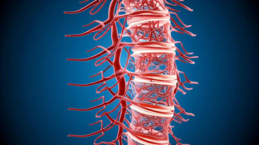
Unraveling Spinal AVMs: A Journey Through Evolving Classification Systems
"Explore how advances in imaging, microsurgery, and endovascular techniques have reshaped the classification of spinal arteriovenous malformations (AVMs) over time."
Spinal arteriovenous malformations (AVMs) present a unique challenge due to the myriad of classification systems proposed over the years. This abundance of systems can lead to confusion, especially when textbooks and research papers seem to contradict each other.
To clarify this complex landscape, we'll explore the evolution of spinal AVM classification, tracing its development through key research and technological advancements. By understanding the historical context, we can better navigate the current classification systems and their practical applications.
This article draws upon English language literature from 1967 to 2015, focusing on landmark papers and updates that have significantly shaped how we understand and categorize these intricate vascular malformations.
From Angiograms to Advanced Imaging: Milestones in AVM Classification

The classification of spinal AVMs has evolved through distinct generations, each marked by advancements in diagnostic and interventional techniques. Here's a look at the key milestones:
- 1971: First description of spinal dural arteriovenous fistulas (AVFs) and intradural AVMs diagnosed by spinal angiography.
- 1986: Second-generation classification based on case reports of intradural perimedullary AVFs treated by microsurgery.
- 1993: Third-generation classification based on case series of intradural perimedullary AVFs treated by endovascular interventions.
- 2002: Fourth-generation classification based on case series of spinal AVMs treated by microsurgery and endovascular interventions.
- 2009 & 2011: Fifth-generation classification based on case series of extradural AVFs treated by microsurgery and endovascular interventions.
Navigating the Future of AVM Classification
The classification of spinal AVMs has been significantly influenced by the evolution of diagnostic imaging, microsurgery, and endovascular treatment. These advancements have allowed for more detailed characterization and understanding of AVM anatomy and hemodynamics.
While various classification systems have been proposed, the third generation, combined with the classification of extradural AVFs, offers a practical framework. Integrating historical context helps to understand the nuances and limitations of each system.
Moving forward, the integration of high-resolution imaging with pathological findings will be crucial for refining AVM classification and treatment strategies. This multidisciplinary approach promises to improve diagnostic accuracy and optimize patient outcomes.
