Surreal illustration of pulmonary amyloidosis in an Australian indiginous population.

Unmasking Pulmonary Amyloidosis: A Mimic of Lung Cancer

"How a Rare Condition Masqueraded as Lung Malignancy in an Indigenous Australian Woman with Sjogren's Syndrome"


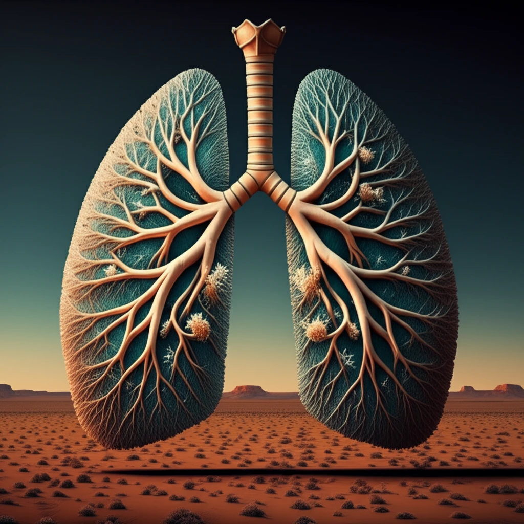
Pulmonary nodules and cysts can arise from many different conditions. Sjogren's syndrome, primarily known for causing dry eyes and mouth, can also affect the lungs. Lung problems linked to Sjogren's typically include interstitial lung disease (ILD) and issues with the trachea and bronchi. In very rare instances, amyloidosis—specifically in the form of nodules and cysts—occurs secondary to Sjogren's syndrome.

This article delves into the case of a 52-year-old Australian Indigenous woman with Sjogren's syndrome, whose initial diagnosis pointed towards metastatic lung cancer due to a concerning nodule found on a PET scan. Further investigation revealed a surprising alternative.

Through this case, we aim to emphasize the diagnostic challenges and the importance of considering less common conditions, even when initial findings suggest something more typical.

The Case: Unveiling the True Diagnosis

Surreal illustration of pulmonary amyloidosis in an Australian indiginous population.

A 52-year-old Australian Indigenous woman, already diagnosed with Sjogren's syndrome a decade prior, presented with a persistent dry cough lasting six months. Aside from this, she reported no other respiratory symptoms or general signs of illness like fever or weight loss. Her history included a teenage smoking habit, and she only took NSAIDs for occasional back pain. Physical examination revealed an enlarged thyroid gland, but was otherwise unremarkable.

Initial lab results showed a normal blood count and liver function, but an elevated ESR (erythrocyte sedimentation rate) and a positive antinuclear antibody (ANA) test. Further tests confirmed the presence of Sjogren's-related antibodies (SS-A and SS-B). A CT scan of her chest revealed multiple lung nodules, including a suspicious mass in the right lower lobe, along with several lung cysts. A PET scan highlighted the nodule with increased activity, raising concerns for malignancy.

  • Bronchoscopy: Initially, a bronchoscopy (a procedure to visualize the airways) found no abnormalities.
  • Biopsy Complication: A CT-guided biopsy of the concerning nodule was attempted, but it led to a pneumothorax (collapsed lung) and yielded inconclusive results.
  • Surgical Revelation: Ultimately, a surgical wedge resection (removal of a small piece of the lung) was performed. Microscopic examination revealed amorphous material that, when stained with Congo red and viewed under polarized light, displayed apple-green birefringence. This distinctive finding confirmed the diagnosis of pulmonary AL amyloidosis.
This meant that the initial suspicion of lung cancer was incorrect. The nodules and cysts were instead due to an unusual buildup of amyloid protein in the lungs, triggered by her underlying Sjogren's syndrome.

Key Takeaways: Amyloidosis and Diagnostic Vigilance

This case underscores the importance of considering rare conditions like pulmonary amyloidosis, especially in patients with autoimmune disorders such as Sjogren's syndrome. While lung nodules and cysts often raise concerns for cancer, this case demonstrates that other possibilities should be thoroughly investigated.

Pulmonary amyloidosis secondary to Sjogren's syndrome is rare and can be difficult to diagnose. The case highlights the diagnostic challenges and the importance of surgical lung biopsy in confirming the diagnosis and excluding other conditions.

This case adds to the growing body of knowledge on the pulmonary manifestations of both amyloidosis and Sjogren's syndrome, reminding clinicians to maintain a broad differential diagnosis and utilize appropriate diagnostic tools to ensure accurate and timely diagnosis, especially in complex presentations.

About this Article -

This article was crafted using a human-AI hybrid and collaborative approach. AI assisted our team with initial drafting, research insights, identifying key questions, and image generation. Our human editors guided topic selection, defined the angle, structured the content, ensured factual accuracy and relevance, refined the tone, and conducted thorough editing to deliver helpful, high-quality information.See our About page for more information.

This article is based on research published under:

DOI-LINK: 10.1016/j.rmcr.2018.11.015, Alternate LINK

Title: Pulmonary Al- Amyloidosis Masquerading As Lung Malignancy In An Australian Indigenous Patient With Sjogren'S Syndrome

Subject: Pulmonary and Respiratory Medicine

Journal: Respiratory Medicine Case Reports

Publisher: Elsevier BV

Authors: Subash Heraganahally, Madeline Digges, Madeleine Haygarth, Kosala Liyanaarachchi, Akash Kalro, Sumit Mehra

Published: 2019-01-01

Everything You Need To Know

1

What is pulmonary amyloidosis, and how is it related to Sjogren's syndrome?

Pulmonary amyloidosis, particularly the AL type, involves the accumulation of amyloid protein within the lung tissue. In the context of Sjogren's syndrome, this can manifest as lung nodules and cysts. The case described demonstrates that while lung cancer is a common concern when nodules are detected, pulmonary amyloidosis should also be considered, especially in individuals with pre-existing autoimmune conditions.

2

Besides dry eyes and mouth, how else can Sjogren's syndrome affect the lungs?

Sjogren's syndrome is an autoimmune disorder primarily known for causing dry eyes and dry mouth. However, it can also affect other organs, including the lungs. Lung manifestations associated with Sjogren's can include interstitial lung disease, and, less commonly, pulmonary amyloidosis. The association with amyloidosis highlights the diverse ways autoimmune diseases can impact different body systems.

3

How is pulmonary AL amyloidosis definitively diagnosed?

The definitive diagnosis of pulmonary AL amyloidosis often requires a tissue biopsy and specific staining techniques. In the case described, a surgical wedge resection was performed, and the tissue sample was stained with Congo red. The presence of apple-green birefringence under polarized light confirmed the presence of amyloid deposits. This microscopic finding is crucial for differentiating amyloidosis from other conditions that may present with similar lung abnormalities.

4

Why did the PET scan initially point toward lung cancer, and what does this say about PET scans?

A PET scan detects areas of increased metabolic activity, which can be indicative of cancer but is not specific to it. In the described case, the PET scan highlighted a lung nodule, raising initial suspicion for malignancy. However, amyloid deposits can also exhibit increased activity on PET scans due to inflammatory processes. Therefore, while a PET scan can be a valuable tool in identifying suspicious lesions, it should be interpreted in conjunction with other diagnostic modalities to avoid misdiagnosis.

5

What is the key takeaway regarding diagnosis when a patient has Sjogren's syndrome and lung nodules?

The case emphasizes the importance of considering rare conditions, such as pulmonary AL amyloidosis, in the differential diagnosis of lung lesions, especially in patients with autoimmune disorders like Sjogren's syndrome. It also highlights the limitations of relying solely on initial imaging findings, such as those from a PET scan, and the necessity of obtaining tissue samples for definitive diagnosis. This diagnostic vigilance can prevent misdiagnosis and ensure appropriate management.

Newsletter Subscribe

Subscribe to get the latest articles and insights directly in your inbox.