Microscopic view of Mycobacterium tuberculosis with a protein structure, symbolizing accurate TB diagnosis.

Unlocking Tuberculosis: How a Protein's Structure Could Revolutionize Diagnosis

"New research sheds light on a unique protein in Mycobacterium tuberculosis, paving the way for more accurate detection of asymptomatic infections and improved disease management."


Tuberculosis (TB) remains a major global health challenge, causing significant mortality and morbidity worldwide. A particularly insidious aspect of TB is its ability to exist in an asymptomatic, or latent, state. It affects about one-third of the world’s population. Between 5 and 10% of those with latent TB will develop active TB during their lifetime, making it essential to develop accurate diagnostic tools for early detection and intervention.

Current diagnostic methods, such as the interferon-gamma release assay (IGRA), have limitations in detecting latent TB, prompting researchers to explore new biomarkers and diagnostic approaches. One promising target is Mycobacterial DNA-binding protein 1 (MDP1), a major cellular protein in Mycobacterium tuberculosis that is highly expressed during the persistent stages of infection. Identifying and understanding the role of this protein could lead to breakthroughs in TB diagnosis.

Recent studies have highlighted MDP1 as a potential diagnostic marker for asymptomatic TB. But, previous research faced a challenge: detecting the antibodies was not specific enough, as they appeared in both infected and non-infected people. Now, a new study is changing how we see MDP1, focusing on its structure to create better diagnostic tools.

Why Protein Structure Matters in TB Diagnosis

Microscopic view of Mycobacterium tuberculosis with a protein structure, symbolizing accurate TB diagnosis.

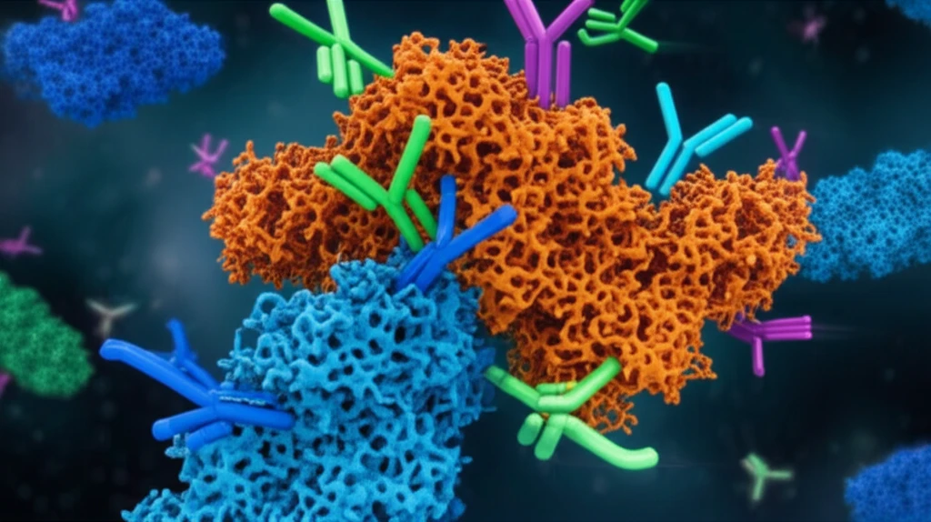
The key to this breakthrough lies in understanding the structure of MDP1. Proteins aren't just simple chains; they fold into complex 3D shapes that determine how they interact with other molecules, including antibodies. The study emphasizes that an antigen's tertiary structure—its unique 3D arrangement—significantly influences antibody recognition. Think of it like a lock and key: the more precise the fit, the stronger the binding and the more accurate the diagnostic test.

Researchers discovered that previous methods of purifying MDP1, which involved acid extraction, likely disrupted its native structure, leading to inaccurate antibody recognition. To overcome this, they developed a refined purification method that preserves MDP1’s natural form. This allowed them to investigate how the protein's structure affects its ability to be recognized by antibodies, with some surprising discoveries:

  • The N-Terminal Half: This part of MDP1 is similar to bacterial histone-like protein HU, known for binding to DNA.
  • The C-Terminal Half: Unlike the N-terminal, this part is believed to be intrinsically disordered, meaning it doesn't have a fixed structure.
  • The Full Picture: The study suggests that both halves of MDP1 are crucial for antibody recognition, highlighting the importance of the protein's overall structure.
By obtaining MDP1 in its native state, the researchers were able to develop an improved IgG-ELISA (enzyme-linked immunosorbent assay) that showed promise in detecting asymptomatic tuberculosis. This refined method demonstrated enhanced specificity and sensitivity, reducing the likelihood of false positives and improving the accuracy of diagnosis.

The Road Ahead: Better TB Detection and Management

This study marks a significant step forward in the fight against tuberculosis. By understanding the crucial role of MDP1’s structure, researchers have paved the way for more accurate diagnostic tools that can detect asymptomatic infections. Earlier and more precise detection of TB could lead to more effective interventions, improved patient outcomes, and reduced transmission rates. While further research and validation are needed, this breakthrough offers hope for better TB control and management worldwide.

About this Article -

This article was crafted using a human-AI hybrid and collaborative approach. AI assisted our team with initial drafting, research insights, identifying key questions, and image generation. Our human editors guided topic selection, defined the angle, structured the content, ensured factual accuracy and relevance, refined the tone, and conducted thorough editing to deliver helpful, high-quality information.See our About page for more information.

Everything You Need To Know

1

What is the significance of Mycobacterial DNA-binding protein 1 (MDP1) in tuberculosis diagnosis?

Mycobacterial DNA-binding protein 1 (MDP1) is a crucial protein in Mycobacterium tuberculosis, the bacterium causing tuberculosis. The article highlights its potential as a diagnostic marker, particularly for asymptomatic tuberculosis. The significance lies in its high expression during the persistent stages of infection. Understanding MDP1's structure allows for the development of more accurate diagnostic tools, improving early detection of tuberculosis.

2

How does the structure of MDP1 influence the accuracy of tuberculosis diagnosis?

The structure of MDP1 plays a critical role in antibody recognition, akin to a lock and key mechanism. The study emphasizes that the tertiary structure of MDP1, its 3D arrangement, significantly influences how antibodies interact with it. When MDP1 is in its native state, the antibodies bind more precisely, leading to stronger binding and more accurate test results. Previous methods disrupted MDP1's native structure, leading to inaccurate antibody recognition and false positives, which is why the new approach focusing on the natural form of MDP1 is vital.

3

What are the key structural features of MDP1 and how do they contribute to antibody recognition?

MDP1 has distinct structural components: the N-terminal half, which is similar to bacterial histone-like protein HU, is known for binding to DNA, and the C-terminal half, which is believed to be intrinsically disordered and lacks a fixed structure. The study indicates that both halves are crucial for antibody recognition. The overall structure is essential for the improved IgG-ELISA test, enhancing both specificity and sensitivity in detecting asymptomatic tuberculosis by allowing the antibodies to recognize the protein correctly, leading to more accurate diagnosis.

4

Why is it important to detect asymptomatic tuberculosis, and how could the new diagnostic tools help?

Detecting asymptomatic tuberculosis is crucial because it affects a significant portion of the world's population. Between 5 and 10% of those with latent TB will develop active TB, which can lead to serious health complications and further spread. The new diagnostic tools, based on a better understanding of MDP1's structure, aim to detect these asymptomatic infections earlier and more accurately. This early detection allows for prompt intervention and treatment, improving patient outcomes and reducing transmission rates, contributing to better TB control and management globally.

5

What are the limitations of existing diagnostic methods for tuberculosis, and how does this new research address them?

Current methods like the interferon-gamma release assay (IGRA) have limitations in detecting latent TB. These methods may not always be sensitive or specific enough. The research addresses these limitations by focusing on the structure of Mycobacterial DNA-binding protein 1 (MDP1). The new approach involves a refined purification method that preserves MDP1’s natural form. By obtaining MDP1 in its native state, researchers developed an improved IgG-ELISA test. This method shows enhanced specificity and sensitivity, reducing false positives and improving diagnostic accuracy, leading to more reliable detection of asymptomatic tuberculosis cases.

Newsletter Subscribe

Subscribe to get the latest articles and insights directly in your inbox.