Magnetic Resonance Elastography (MRE) image of a liver with color-coded stiffness.

Unlocking the Secrets of Soft Tissue: How Advanced Imaging Can Detect Hidden Health Issues

"Magnetic Resonance Elastography (MRE) is revolutionizing medical diagnostics by providing a more accurate and nuanced understanding of tissue stiffness, potentially leading to earlier and more precise disease detection."


Imagine being able to 'feel' what's going on inside your body without any invasive procedures. That's the promise of Magnetic Resonance Elastography (MRE), a cutting-edge medical imaging technique that's offering doctors a new way to assess the health of your soft tissues. Traditionally, methods like palpation (physical examination by touch) and even standard imaging techniques often miss subtle but crucial changes within the body. MRE steps in to fill that gap, providing a non-invasive method to measure tissue stiffness, which can be an indicator of underlying health issues.

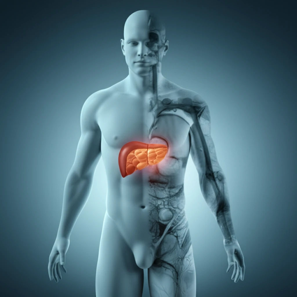
The stiffness of your tissues isn't just a random characteristic; it's a key biomechanical property that can signal the presence of diseases like fibrosis, cancer, and other disorders long before they're detectable through conventional means. Think of it like this: a healthy liver feels different than a liver affected by cirrhosis. MRE captures these differences with incredible precision, translating them into detailed images that help doctors make more informed diagnoses.

This article delves into the science behind MRE, exploring how it works, what it can reveal, and why it's poised to become an indispensable tool in modern medicine. We'll break down the technical jargon, explain the clinical implications, and show you how this innovative technology is changing the landscape of diagnostics and treatment.

MRE: The Science of 'Feeling' Inside

Magnetic Resonance Elastography (MRE) image of a liver with color-coded stiffness.

At its core, MRE is a sophisticated imaging method that combines magnetic resonance imaging (MRI) with principles of elasticity. Unlike standard MRI, which primarily visualizes anatomical structures, MRE assesses the mechanical properties of tissues by measuring how they respond to vibrations. Here's the breakdown:

A device generates tiny, controlled vibrations that pass through the targeted tissue. These vibrations create 'waves' within the tissue.

  • The MRE machine tracks the propagation of these waves using modified MRI sequences.
  • Sophisticated computer algorithms analyze the wave patterns, calculating tissue stiffness based on how quickly and easily the waves travel.
  • The results are displayed as a color-coded map, called an elastogram, showing areas of varying stiffness. Stiffer areas might indicate disease, while softer areas suggest healthy tissue or other conditions.
One of the key advantages of MRE is its ability to quantify stiffness, not just visualize it. This provides doctors with objective data to support their diagnoses and monitor treatment effectiveness. MRE also works non-invasively, eliminating the risks associated with biopsies or other invasive procedures.

The Future of MRE: Broader Applications and Personalized Medicine

As research continues, the potential applications of MRE are expanding beyond the liver to include the breast, heart, and even the brain. The ability to non-invasively assess tissue health has profound implications for early disease detection, personalized treatment planning, and monitoring the effectiveness of therapies. By unlocking the secrets hidden within our soft tissues, MRE is paving the way for a future of more proactive and precise healthcare.

About this Article -

This article was crafted using a human-AI hybrid and collaborative approach. AI assisted our team with initial drafting, research insights, identifying key questions, and image generation. Our human editors guided topic selection, defined the angle, structured the content, ensured factual accuracy and relevance, refined the tone, and conducted thorough editing to deliver helpful, high-quality information.See our About page for more information.

Everything You Need To Know

1

What exactly is Magnetic Resonance Elastography (MRE)?

Magnetic Resonance Elastography (MRE) is a cutting-edge medical imaging technique designed to measure the stiffness of soft tissues within the body. Unlike traditional methods, MRE uses a combination of magnetic resonance imaging (MRI) and principles of elasticity to provide detailed information about tissue health. A device generates tiny vibrations that pass through the tissue, and the MRE machine tracks the propagation of these waves. Sophisticated computer algorithms then analyze the wave patterns to calculate tissue stiffness, creating a color-coded map called an elastogram.

2

Why is Magnetic Resonance Elastography (MRE) considered important?

MRE is important because it allows for earlier and more accurate disease detection. The stiffness of soft tissues can be an indicator of underlying health issues, such as fibrosis, cancer, and other disorders, often before they are detectable through conventional methods. By quantifying tissue stiffness non-invasively, MRE provides doctors with objective data to support diagnoses and monitor treatment effectiveness. This capability is crucial for proactive healthcare.

3

What are the implications of using Magnetic Resonance Elastography (MRE) in healthcare?

The implications of MRE are far-reaching, particularly for early disease detection and personalized medicine. By providing detailed information about tissue stiffness, MRE enables doctors to identify diseases at earlier stages, potentially leading to more effective treatments. The technology’s non-invasive nature eliminates risks associated with biopsies. As research expands, MRE's applications are broadening to include the breast, heart, and even the brain, further enhancing its potential to transform diagnostics and treatment.

4

How does Magnetic Resonance Elastography (MRE) work?

MRE works by combining magnetic resonance imaging (MRI) with principles of elasticity. A device generates tiny, controlled vibrations that pass through the targeted tissue. These vibrations create 'waves' within the tissue. The MRE machine tracks the propagation of these waves using modified MRI sequences. Sophisticated computer algorithms analyze the wave patterns, calculating tissue stiffness based on how quickly and easily the waves travel. The results are displayed as a color-coded map, called an elastogram, showing areas of varying stiffness.

5

What is an elastogram in the context of Magnetic Resonance Elastography (MRE)?

The elastogram is a color-coded map generated by Magnetic Resonance Elastography (MRE) that displays areas of varying tissue stiffness. Stiffer areas might indicate disease, while softer areas suggest healthy tissue or other conditions. The elastogram provides doctors with a visual representation of tissue stiffness, enabling them to make more informed diagnoses and monitor the effectiveness of treatments. This is a key feature that distinguishes MRE from traditional imaging methods.

Newsletter Subscribe

Subscribe to get the latest articles and insights directly in your inbox.