Unlocking the Secrets of P63-Associated Disorders: How New Research Offers Hope for Skin Health
"A Deep Dive into the Genetic Mechanisms Behind Rare Syndromes and Potential Breakthroughs in Treatment."
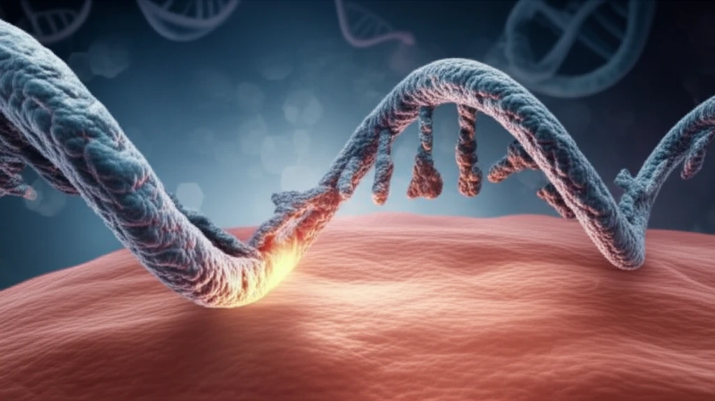
The p53 family member p63 plays a vital role in gene expression within stratified epithelia, which includes the epidermis. One of its major functions is sustaining mechanical resistance by positively regulating several epidermal genes involved in cell-matrix adhesion and cell-cell adhesion. When p63 is deficient, it can lead to severe defects in stratified epithelia and their derivatives, impacting overall health.
Heterozygous mutations in the p63 gene in humans can lead to at least five rare, related syndromes with overlapping features. These include ectodermal dysplasia, cleft lip/palate, and limb malformations. Among these disorders, one notable condition is Ectrodactyly, ectodermal dysplasia, and cleft lip/palate syndrome 3 (EEC). This syndrome is often characterized by central reduction defects of the hands and feet, associated with cleft lip and/or palate, and is frequently caused by mutations in the DNA-binding domain that disrupt p63's binding ability.
In contrast, Ankyloblepharon-ectodermal defects-cleft lip/palate (AEC) syndrome presents with severe skin symptoms such as congenital erythroderma, skin fragility, atrophy, palmo-plantar hyperkeratosis, and extensive, long-lasting erosive lesions. The mechanisms behind AEC skin erosions are not fully understood, standard care focuses on minimizing trauma and preventing infections.
How Does Mutant p63 Lead to Skin Problems?
To understand the molecular basis of AEC syndrome, researchers generated a knock-in mouse model that expresses a mutation in the leucine 514 to phenylalanine (L514F). This mutation is significant because leucine 514 is highly conserved and found mutated in phenylalanine, valine, or serine in AEC patients. The AEC mouse model closely resembles the human disorder, presenting with cleft palate, skin atrophy, hair and tooth defects, but without ectrodactyly.
- Skin atrophy and delayed wound healing are linked to a reduced number of epidermal stem cells.
- This reduction is caused by compromised fibroblast growth factor receptor signaling and downregulation of Fgfr2 and Fgfr3, which are p63 target genes.
- A defective stem cell compartment is also observed in human skin affected by AEC syndrome.
- Desmosomes, essential cell junctions for mechanical resistance in the epidermis, are weakened in AEC syndrome.
- This is due to reduced gene expression of p63 direct target genes like Dsc3, Dsg1, and Dsp.
The Role of TSLP in Skin Inflammation and Autoimmunity
Skin erosions trigger local and systemic inflammatory responses that depend on TSLP, an IL-7-like cytokine. TSLP is produced by keratinocytes and released in response to epidermal barrier failure. In human and mouse skin, TSLP is highly expressed in acute and chronic lesions associated with atopic dermatitis and Netherton syndrome. TSLP has wide-ranging effects on cell lineages, including immune cells, and drives Th2-mediated inflammation. In AEC mice, Tslp circulates in the blood, causing expansion of pre-B cells and leading to an autoimmune B-cell proliferative disorder. Ablating Tslp in the epidermis of AEC mice reduces skin and systemic inflammation, rescues B-cell differentiation, and reduces mortality.