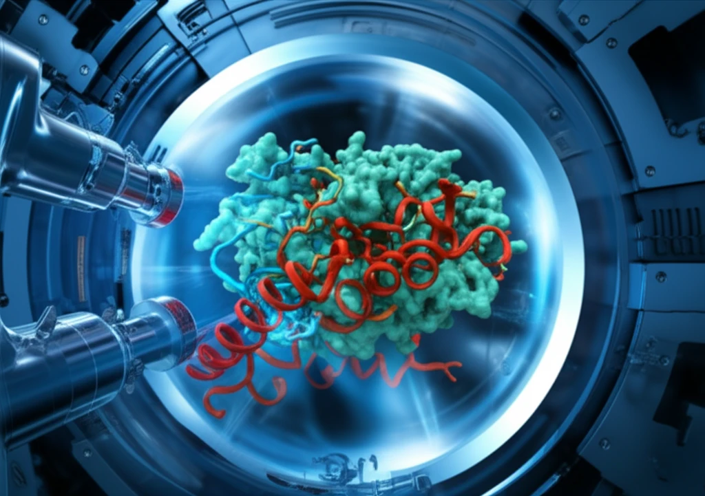
Unlocking the Secrets of Life: How Cryo-Electron Microscopy is Revolutionizing Structural Biology
"Discover how cryo-EM is changing our understanding of proteins and their dynamic behaviors, offering new insights into disease and potential treatments."
The 2017 Nobel Prize in Chemistry recognized the pioneers of cryo-electron microscopy (cryo-EM), a method that has reshaped how we visualize the intricate world of proteins. Traditionally, X-ray crystallography and NMR spectroscopy were the dominant techniques for determining protein structures. However, thanks to decades of dedicated effort by numerous scientists, cryo-EM has emerged as a powerful third option [1].
Cryo-EM distinguishes itself by directly imaging protein samples using an electron microscope. This eliminates the need for crystallization or isotopic labeling, allowing researchers to study proteins in a more native-like state. Furthermore, cryo-EM requires only minute amounts of sample and is particularly well-suited for analyzing large protein complexes [1].
Recent advancements have extended cryo-EM's capabilities to dynamic structural analysis, providing insights into how proteins change their conformations in response to various stimuli. Unlike other methods, cryo-EM imposes minimal spatial constraints, making it ideal for studying the dynamic behavior of large protein complexes and capturing structural changes during ligand binding or protein-protein interactions. By analyzing the proportions of different dynamic states, researchers can even glean information about the kinetics of molecular processes.
The Principles of Cryo-Electron Microscopy: How Does It Work?

Biological samples, like proteins, typically rely on water to maintain their shape and function. However, electron microscopes operate under high vacuum conditions (below 10⁻⁵ Pa) to allow electrons to travel freely. Introducing a hydrated biological sample into this environment would cause it to dry out and collapse, losing its native structure and function. Additionally, biological molecules are primarily composed of light elements like carbon, which scatter electrons weakly [2].
- Traditional Approaches: Earlier electron microscopy of biological samples relied on techniques like negative staining, where samples were embedded in heavy metal salts like uranyl acetate (Figure 2a). This allowed for visualization based on differences in electron scattering between the heavy metal stain and the sample [3].
- Limitations of Negative Staining: While useful, negative staining only provided information about the sample's outline and lacked the ability to resolve internal structural details at high resolution, preventing the determination of atomic coordinates.
- Cryo-EM's Solution: Cryo-EM overcomes these limitations by employing a technique called cryo-protection, developed by Jacques Dubochet, one of the Nobel laureates. This involves rapidly freezing the biological sample in a thin film of amorphous ice, effectively trapping it in a near-native state (Figure 2b). This vitrified sample is then loaded into the electron microscope and imaged [4].
Looking Ahead: The Future of Cryo-EM
This review has highlighted the power of single-particle cryo-EM as a leading method for protein structure determination. Its minimal sample requirements and ability to study proteins in near-native conditions pave the way for exciting future applications. Besides single-particle analysis, cryo-electron tomography and subtomogram averaging offer alternative approaches, enabling the study of heterogeneous samples and proteins within cellular contexts, although with slightly reduced resolution. As cryo-EM technology continues to advance, expect even more exciting developments and broader adoption across various scientific disciplines.
