Collagen strands intertwined with DNA, highlighting collagen degradation.

Unlocking the Secrets of Collagen: How New Imaging Tech Could Revolutionize Disease Detection

"Cutting-edge peptide hybridization techniques offer a glimpse into collagen breakdown, promising earlier diagnosis and more effective treatment strategies."


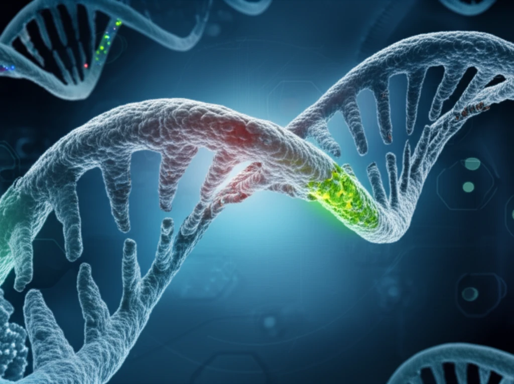
The extracellular matrix (ECM), particularly its most abundant component collagen, plays a critical role in tissue health. When collagen breaks down, it signals various diseases, including cancer, cardiovascular issues, and organ fibrosis. Current methods for detecting this degradation are limited, hindering early diagnosis and effective treatment strategies. The need for advanced tools to monitor collagen breakdown is paramount.

Traditional methods rely on synthetic hydrogels or cells with reporter genes, but these often fail to replicate the natural ECM environment or are incompatible with primary cells. While techniques like second harmonic generation (SHG) microscopy can visualize the fibrillar structure of ECM, they lack the sensitivity needed to detect subtle changes at the molecular level.

Enter the collagen hybridizing peptide (CHP), a revolutionary tool that specifically visualizes protease-degraded collagen in vitro and in vivo. This technology offers new possibilities for understanding ECM pathobiology and developing targeted therapies.

Visualizing Collagen Breakdown: A New Frontier in Disease Detection

Collagen strands intertwined with DNA, highlighting collagen degradation.

Researchers have developed a groundbreaking technique using collagen hybridizing peptides (CHPs) to visualize collagen proteolysis. These peptides, designed to bind to unfolded collagen chains, offer a direct way to observe collagen degradation, a hallmark of many diseases. By fluorescently labeling CHPs, scientists can track collagen breakdown in real-time, providing unprecedented insights into disease progression.

A key innovation involves modifying the CHP sequence with (2S,4S)-4-fluoroproline (f), preventing self-assembly into triple helices and enhancing its ability to target degraded collagen in vivo. This modified CHP, tagged with a near-infrared fluorophore, enables imaging and quantification of osteolytic bone lesions in mouse models of multiple myeloma, showcasing its potential for early disease detection and personalized treatment.

  • Enhanced Specificity: CHP selectively targets degraded collagen, minimizing off-target effects.
  • Real-Time Visualization: Fluorescent labeling allows for dynamic monitoring of collagen breakdown.
  • Non-Invasive Imaging: Modified CHPs enable in vivo imaging, reducing the need for invasive procedures.
  • Versatile Applications: Suitable for a range of diseases, from cancer to arthritis, offering broad diagnostic potential.
The CHP-mediated imaging correlates with conventional techniques like micro-CT scans but offers a more versatile approach for assessing bone lesions. This technology promises to revolutionize our understanding of ECM biology and facilitate the development of clinical molecular imaging.

The Future of Collagen-Targeted Therapies

The development of collagen hybridizing peptides (CHPs) marks a significant leap forward in disease diagnostics and treatment. By providing a direct and specific method for visualizing collagen degradation, this technology overcomes the limitations of traditional approaches and opens new avenues for early disease detection.

Looking ahead, CHPs hold promise for a wide range of biomedical applications, from studying ECM biology to developing clinical molecular imaging techniques. Their ability to provide real-time insights into collagen breakdown could revolutionize our approach to personalized medicine.

Further research and development in this area could lead to more effective therapies and improved patient outcomes. As we continue to unlock the secrets of collagen, we move closer to a future where diseases are detected and treated earlier, leading to healthier and longer lives.

About this Article -

This article was crafted using a human-AI hybrid and collaborative approach. AI assisted our team with initial drafting, research insights, identifying key questions, and image generation. Our human editors guided topic selection, defined the angle, structured the content, ensured factual accuracy and relevance, refined the tone, and conducted thorough editing to deliver helpful, high-quality information.See our About page for more information.

This article is based on research published under:

DOI-LINK: 10.1016/j.biomaterials.2018.08.039, Alternate LINK

Title: Visualizing Collagen Proteolysis By Peptide Hybridization: From 3D Cell Culture To In Vivo Imaging

Subject: Mechanics of Materials

Journal: Biomaterials

Publisher: Elsevier BV

Authors: Lucas L. Bennink, Yang Li, Bumjin Kim, Ik Jae Shin, Boi Hoa San, Maurizio Zangari, Donghoon Yoon, S.Michael Yu

Published: 2018-11-01

Everything You Need To Know

1

How do Collagen Hybridizing Peptides (CHPs) actually work to show collagen breakdown?

Collagen Hybridizing Peptides (CHPs) are designed to bind specifically to unfolded collagen chains, which are exposed when collagen is broken down. By fluorescently labeling these CHPs, scientists can directly visualize and track collagen degradation in real-time. This targeted approach minimizes off-target effects and offers a dynamic way to monitor collagen breakdown, providing unprecedented insights into disease progression. This contrasts with older methods that struggled to replicate natural extracellular matrix (ECM) environments.

2

What are the drawbacks of old collagen detection techniques, and how do Collagen Hybridizing Peptides (CHPs) improve upon them?

Traditional methods for detecting collagen degradation, such as synthetic hydrogels, cells with reporter genes, and even second harmonic generation (SHG) microscopy, have limitations. Synthetic hydrogels and reporter genes often fail to accurately mimic the natural ECM environment or are incompatible with primary cells. SHG microscopy can visualize the fibrillar structure of the ECM, but lacks the sensitivity needed to detect subtle molecular-level changes associated with early collagen degradation. Collagen Hybridizing Peptides (CHPs) overcome these limitations by directly targeting and visualizing degraded collagen with high specificity and sensitivity.

3

Why is modifying the Collagen Hybridizing Peptide (CHP) sequence with (2S,4S)-4-fluoroproline (f) so important?

The modification of the Collagen Hybridizing Peptide (CHP) sequence with (2S,4S)-4-fluoroproline (f) is crucial because it prevents the CHP from self-assembling into triple helices. This enhances the modified CHP's ability to target degraded collagen in vivo. Tagging this modified CHP with a near-infrared fluorophore allows for in vivo imaging and quantification of osteolytic bone lesions, as demonstrated in mouse models of multiple myeloma. Without this modification, the CHP's effectiveness in targeting degraded collagen within a living organism would be significantly reduced.

4

How could visualizing collagen breakdown using Collagen Hybridizing Peptides (CHPs) change how we treat diseases in the future?

Visualizing collagen degradation using Collagen Hybridizing Peptides (CHPs) has broad implications for personalized medicine. By enabling early and precise detection of diseases like cancer, cardiovascular issues, and arthritis, CHPs can facilitate the development of targeted therapies tailored to individual patients. For instance, the ability to image and quantify osteolytic bone lesions in multiple myeloma mouse models demonstrates the potential for early disease detection and personalized treatment strategies based on real-time monitoring of collagen breakdown.

5

Why is visualizing collagen degradation with Collagen Hybridizing Peptides (CHPs) such a big step forward in finding and treating diseases?

Collagen Hybridizing Peptides (CHPs) represent a new approach to disease diagnostics and treatment because they provide a direct and specific method for visualizing collagen degradation. Unlike traditional methods that often lack sensitivity or fail to replicate the natural ECM environment, CHPs can target degraded collagen in vivo, offering real-time visualization and quantification. This opens new avenues for early disease detection, personalized medicine, and the development of collagen-targeted therapies, potentially revolutionizing our understanding of ECM biology and clinical molecular imaging. The advantage is that CHPs can also correlate with conventional techniques like micro-CT scans but offer a more versatile approach for assessing bone lesions. What is missing is the study of direct effects of the peptides in humans.

Newsletter Subscribe

Subscribe to get the latest articles and insights directly in your inbox.