Illustration of a glowing centrosome inside a dividing cell.

Unlocking the Secrets of Cell Division: How Scientists are Mapping the Centrosome

"New research provides an unprecedented look at the architecture of the centrosome, offering potential breakthroughs in understanding cancer, ciliopathies, and more."


Cell division is a fundamental process of life, and at the heart of it lies a tiny structure called the centrosome. Centrosomes ensure that when cells divide, each new cell gets the right number of chromosomes. When things go wrong with centrosomes, it can lead to a host of diseases, including cancer, microcephaly (abnormally small head), and ciliopathies (diseases related to defective cilia).

For years, scientists have been trying to understand exactly how centrosomes are built and how they work. Key to this understanding is knowing the precise amounts and arrangement of the proteins that make up the centrosome. It's like understanding how a car engine works – you need to know all the parts and how they fit together.

Now, a team of researchers has made a significant leap forward. They've developed advanced techniques to measure the quantity and organization of key proteins within the human centrosome. This detailed map provides new insights into the centrosome's architecture and opens doors for future studies on cell division and related diseases.

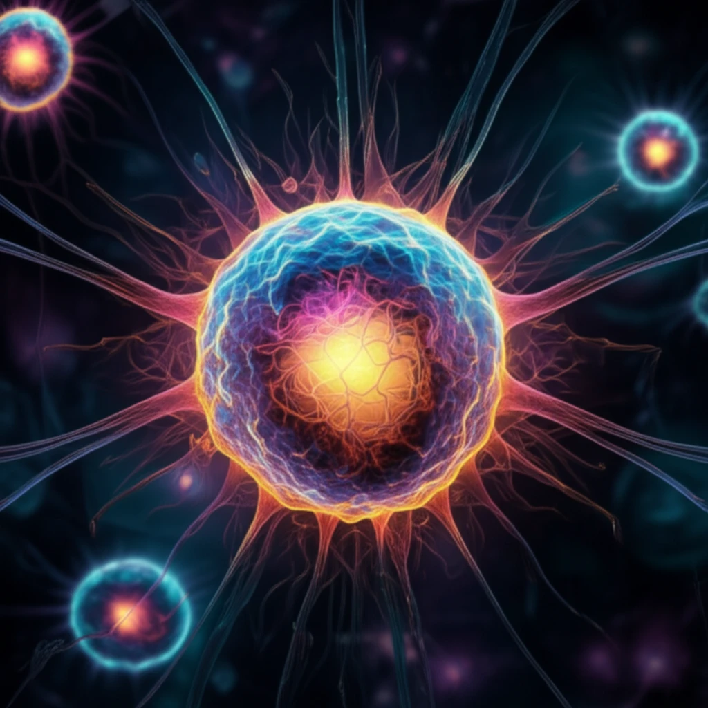
Centrosomes: The Cell's Control Centers

Illustration of a glowing centrosome inside a dividing cell.

Centrosomes are the primary microtubule-organizing centers (MTOCs) in animal cells. Typically, they consist of two centrioles surrounded by a cloud of proteins known as the pericentriolar material (PCM). This intricate structure plays a crucial role in:

While the structure of centrosomes has been known for many years, understanding the exact quantities and arrangement of their protein components has remained a challenge. Recent advances in proteomics and imaging are changing that. Here are some of the roles of centrosomes in key cellular functions:

  • Cell Division: Ensuring accurate chromosome segregation during cell division.
  • Cellular Structure: Giving the cell its shape and internal organization.
  • Motility: Helping cells move and migrate.
  • Cilia Formation: Serving as the foundation for cilia, which are essential for many biological functions.
When centrosomes don't function correctly, the consequences can be severe. Abnormal centrosome numbers or structures have been linked to various diseases, highlighting the importance of understanding these cellular components.

A Quantitative Leap in Understanding

This new research combines two powerful techniques to gain unprecedented quantitative data about centrosomes. The first, targeted proteomics, uses a method called selected reaction monitoring (SRM) to precisely measure the amounts of specific proteins in cell lysates and purified centrosomes. The second approach involves tagging proteins at their natural locations within the cell using a green fluorescent protein (EGFP), allowing researchers to visualize and count the proteins using fluorescence microscopy.

About this Article -

This article was crafted using a human-AI hybrid and collaborative approach. AI assisted our team with initial drafting, research insights, identifying key questions, and image generation. Our human editors guided topic selection, defined the angle, structured the content, ensured factual accuracy and relevance, refined the tone, and conducted thorough editing to deliver helpful, high-quality information.See our About page for more information.

This article is based on research published under:

DOI-LINK: 10.15252/embj.201694462, Alternate LINK

Title: Quantitative Analysis Of Human Centrosome Architecture By Targeted Proteomics And Fluorescence Imaging

Subject: General Immunology and Microbiology

Journal: The EMBO Journal

Publisher: EMBO

Authors: Manuel Bauer, Fabien Cubizolles, Alexander Schmidt, Erich A Nigg

Published: 2016-08-18

Everything You Need To Know

1

What exactly is a centrosome, and why is it important in cell biology?

The centrosome is a key structure within animal cells, acting as the main microtubule-organizing center (MTOC). It is typically composed of two centrioles surrounded by a cloud of proteins known as the pericentriolar material (PCM). The centrosome is crucial for cell division, ensuring each new cell receives the correct number of chromosomes. Dysfunction of the centrosome can lead to diseases like cancer and ciliopathies.

2

Why is it so important to quantitatively analyze the centrosome?

The quantitative analysis of the centrosome provides a detailed understanding of the amounts and organization of its protein components. This is significant because knowing precisely how these proteins are arranged and how much of each exists within the centrosome is vital for understanding its function, similar to understanding the parts of a car engine and how they fit together. This knowledge can help in treating diseases linked to centrosome dysfunction.

3

What key functions do centrosomes perform in cells, and what happens if they don't work correctly?

Centrosomes play several critical roles including ensuring accurate chromosome segregation during cell division, giving the cell its shape and internal organization, helping cells move and migrate, and serving as the foundation for cilia formation, which are essential for many biological functions. If centrosomes don't function correctly, it can lead to severe consequences such as abnormal centrosome numbers or structures, which have been linked to various diseases.

4

How does targeted proteomics help scientists study centrosomes?

Targeted proteomics, particularly selected reaction monitoring (SRM), is used to precisely measure the amounts of specific proteins in cell lysates and purified centrosomes. This technique allows researchers to quantify the protein components of the centrosome accurately. This is important because understanding the precise quantities of these proteins is essential for understanding centrosome function and how disruptions can lead to disease.

5

How does using green fluorescent protein (EGFP) help in studying centrosomes?

The use of green fluorescent protein (EGFP) involves tagging proteins at their natural locations within the cell. This allows researchers to visualize and count the proteins using fluorescence microscopy. This visualization is essential for understanding the spatial arrangement and organization of proteins within the centrosome. Combining this with quantitative data from proteomics offers a comprehensive view of the centrosome's architecture.

Newsletter Subscribe

Subscribe to get the latest articles and insights directly in your inbox.