Fragile brain aneurysm glowing within blood vessels, symbolizing the need for precise monitoring.

Unlocking the Secrets of Brain Aneurysms: How Vessel Wall Imaging is Changing Everything

"New insights into intracranial aneurysms reveal the critical role of vessel wall imaging in predicting rupture risk and improving treatment strategies."


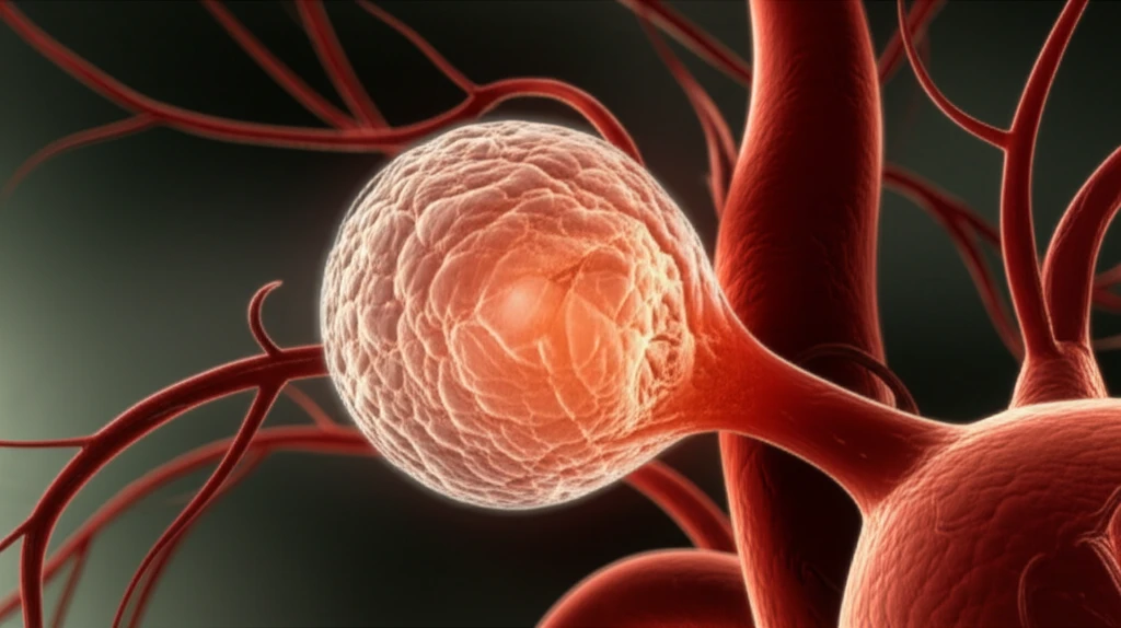
Intracranial aneurysms (IAs), often described as bulges in the brain's blood vessels, are more common than many realize, affecting a significant portion of the population. While some remain stable and asymptomatic, others can rupture, leading to severe complications such as hemorrhagic stroke, a leading cause of death and disability worldwide. This silent threat underscores the importance of understanding the factors that contribute to aneurysm formation and rupture.

For years, scientists have been working tirelessly to unravel the complex mechanisms behind IAs. Inflammation has emerged as a key player, with research showing its involvement in both the development and rupture of these aneurysms. Despite advances in treatment options, managing unruptured IAs remains a challenge, highlighting the need for better diagnostic tools and risk assessment strategies.

Enter magnetic resonance vessel wall imaging (MR-VWI), a cutting-edge technique that allows doctors to visualize the walls of blood vessels in unprecedented detail. By providing a clear picture of vessel wall characteristics, MR-VWI offers valuable insights into the health and stability of IAs. Recent studies have explored the potential of MR-VWI to identify aneurysms at high risk of rupture, paving the way for more targeted and effective interventions.

Decoding Vessel Wall Imaging: What Does It Tell Us About Aneurysm Risk?

Fragile brain aneurysm glowing within blood vessels, symbolizing the need for precise monitoring.

A groundbreaking study featured in Stroke journal shed light on the importance of vessel wall imaging in assessing IA rupture risk. Researchers used MR-VWI to examine unruptured IAs, followed by a detailed histopathological analysis. The results revealed key differences between aneurysms that showed enhancement on MR-VWI and those that did not.

Aneurysms with vessel wall enhancement typically exhibited:

  • Increased wall thickness.
  • Signs of atherosclerosis (plaque buildup).
  • Neovascularization (formation of new blood vessels).
  • Macrophage infiltration (immune cells associated with inflammation).
In contrast, aneurysms without enhancement displayed thinner walls and lacked the cellular density seen in enhanced aneurysms. This suggests that vessel wall enhancement on MR-VWI is indicative of an active inflammatory process within the aneurysm, potentially weakening the vessel wall and increasing the risk of rupture.

The Future of Aneurysm Management: A Personalized Approach

The insights gained from MR-VWI have the potential to revolutionize how we manage intracranial aneurysms. By identifying high-risk aneurysms early on, doctors can develop personalized treatment plans to prevent rupture and its devastating consequences. Further research is needed to fully understand the complex interplay between vessel wall inflammation, aneurysm characteristics, and rupture risk. However, one thing is clear: vessel wall imaging is a game-changer in the fight against brain aneurysms.

About this Article -

This article was crafted using a human-AI hybrid and collaborative approach. AI assisted our team with initial drafting, research insights, identifying key questions, and image generation. Our human editors guided topic selection, defined the angle, structured the content, ensured factual accuracy and relevance, refined the tone, and conducted thorough editing to deliver helpful, high-quality information.See our About page for more information.

This article is based on research published under:

DOI-LINK: 10.1161/strokeaha.118.023701, Alternate LINK

Title: Magnetic Resonance Vessel Wall Imaging In Human Intracranial Aneurysms

Subject: Advanced and Specialized Nursing

Journal: Stroke

Publisher: Ovid Technologies (Wolters Kluwer Health)

Authors: Joseph S. Hudson, Mario Zanaty, Daichi Nakagawa, David K. Kung, Pascal Jabbour, Edgar A. Samaniego, David Hasan

Published: 2019-01-01

Everything You Need To Know

1

What are intracranial aneurysms, and why should I be concerned about them?

Intracranial aneurysms (IAs) are bulges in the brain's blood vessels, affecting a significant portion of the population. While some IAs remain stable, others can rupture, leading to hemorrhagic stroke, a leading cause of death and disability. Recognizing the prevalence and potential severity of IAs highlights the importance of early detection and effective management strategies, which vessel wall imaging is designed to improve.

2

What is magnetic resonance vessel wall imaging (MR-VWI), and how does it work?

Magnetic resonance vessel wall imaging (MR-VWI) is a cutting-edge technique that provides detailed visualization of blood vessel walls. This allows doctors to assess the health and stability of intracranial aneurysms (IAs). By identifying specific characteristics within the vessel walls, like inflammation and plaque buildup, MR-VWI helps determine the risk of aneurysm rupture. This provides valuable insights that were previously unavailable, changing how IAs are managed.

3

Why is vessel wall enhancement on MR-VWI important for understanding aneurysm risk?

Vessel wall enhancement on magnetic resonance vessel wall imaging (MR-VWI) is significant because it indicates an active inflammatory process within the aneurysm. This inflammation weakens the vessel wall, increasing the risk of rupture. The presence of increased wall thickness, atherosclerosis, neovascularization, and macrophage infiltration, as revealed by MR-VWI, are all signs of this inflammatory process. These findings help doctors identify high-risk intracranial aneurysms and implement timely interventions.

4

How can the insights from MR-VWI change the way intracranial aneurysms are managed?

MR-VWI helps doctors identify high-risk intracranial aneurysms (IAs) early on. By understanding the vessel wall characteristics, such as the presence of inflammation, plaque buildup, and other indicators, doctors can develop personalized treatment plans. This targeted approach aims to prevent aneurysm rupture and the severe consequences of hemorrhagic stroke. This approach improves patient outcomes and reduces the risk of disability and death.

5

How are intracranial aneurysms treated, and what role does vessel wall imaging play in their management?

Intracranial aneurysms (IAs) are managed by understanding their rupture risk and using appropriate interventions. MR-VWI is a key tool in this process. Identifying high-risk IAs through MR-VWI allows for targeted treatments, such as endovascular coiling or surgical clipping, to prevent rupture. Regular monitoring and lifestyle adjustments may also be part of a personalized treatment plan, all aimed at minimizing the risk associated with these aneurysms. Future research aims to further refine the understanding of IAs and improve treatment strategies.

Newsletter Subscribe

Subscribe to get the latest articles and insights directly in your inbox.