Unlocking the Secrets: How MRI Scans Revolutionize Diagnosis of MRKH Syndrome
"A Deep Dive into Magnetic Resonance Imaging for Unveiling Müllerian Remnants and Improving Outcomes in Mayer-Rokitansky-Küster-Hauser Syndrome"
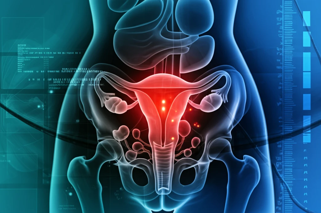
Mayer-Rokitansky-Küster-Hauser (MRKH) syndrome is a congenital condition affecting approximately 1 in 4000 females, characterized primarily by the absence or underdevelopment of the uterus and the upper two-thirds of the vagina. This can lead to significant physical and emotional challenges for those affected, often prompting them to seek gynecological and surgical interventions.
Historically, diagnosing MRKH syndrome and planning surgical treatments have relied on methods like laparoscopy and ultrasonography (USG). Laparoscopy, while providing direct visualization, is an invasive procedure. Ultrasonography, a non-invasive alternative, often falls short in delivering comprehensive anatomical details. This is where Magnetic Resonance Imaging (MRI) steps in to bridge the gap, offering a detailed, non-invasive method for evaluating the condition.
MRI's ability to produce high-resolution images of soft tissues makes it an invaluable tool for assessing Müllerian remnants – the residual tissues from the underdeveloped Müllerian ducts, which would normally form the uterus, fallopian tubes, cervix and upper vagina. By understanding the precise anatomical features of these remnants, as well as any associated anomalies, clinicians can tailor surgical approaches to improve outcomes and quality of life for individuals with MRKH syndrome.
What Does MRI Reveal About Müllerian Remnants in MRKH Syndrome?
A recent study published in the Korean Journal of Radiology delved into the MRI findings of young women suspected of having MRKH syndrome. The research focused on analyzing Müllerian remnants, vagina, ovaries, and other associated findings to establish a clearer understanding of the syndrome's anatomical variations. Fifteen young females underwent multiplanar T2- and transverse T1-weighted MRI scans to facilitate this detailed evaluation.
- Bilateral Uterine Buds: All participants displayed bilateral uterine buds located in the pelvic cavity. These buds, representing underdeveloped uterine structures, had an average long-axis diameter of 2.64 ± 0.65 cm.
- Fibrous Band-Like Structures: In every case, the uterine buds were connected by fibrous band-like structures. These bands converged at the midline or a paramedian triangular soft tissue lying above the bladder dome.
- Vaginal Development: The lower one-third of the vagina was identified in 14 out of 15 cases, providing insights into the extent of vaginal development.
- Ovarian Position: Fourteen cases showed bilateral normal ovaries located near the uterine buds, confirming the presence of functional ovaries, a typical characteristic of MRKH syndrome.
- Associated Anomalies: The study also noted associated anomalies such as a unilateral pelvic kidney, unilateral renal agenesis, mild scoliosis, and lumbar sacralization in some participants.
Why MRI is Becoming the Preferred Diagnostic Tool
The detailed anatomical insights provided by MRI are revolutionizing the diagnostic and surgical approaches to MRKH syndrome. Unlike more invasive methods, MRI offers a non-invasive way to visualize Müllerian remnants and associated anomalies, leading to more informed treatment decisions and improved patient outcomes. As MRI technology continues to advance, it will likely become the gold standard for diagnosing and managing MRKH syndrome, offering hope and improved quality of life for those affected.