Surreal illustration of a giant intracranial aneurysm within a complex brain, symbolizing advanced treatment options.

Unlocking the Secrets: A Modern Guide to Treating Giant Brain Aneurysms

"Navigate the complexities of giant intracranial aneurysms with our comprehensive guide to microsurgery and modern treatment options."


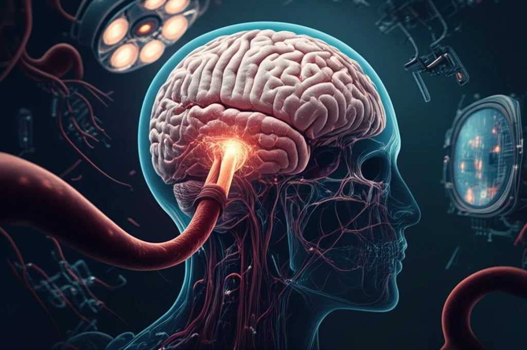
Giant intracranial aneurysms (GIAs), defined as those larger than 25 mm, present a unique and often daunting challenge in neurosurgery. These aneurysms, which can be saccular, fusiform, or dolichoectatic, account for approximately 5% of all cerebral aneurysms. Unlike their smaller counterparts, GIAs tend to appear later in life and are more prevalent in women. Their complex nature stems from wide, atherosclerotic necks, involvement of critical brain branches, the presence of thrombus within the aneurysm, calcified walls, and intricate anatomical structures.

The prognosis for untreated GIAs is grim, with mortality rates soaring to 68% within two years and 85% within five years of diagnosis. These lesions can manifest through various symptoms, including hemorrhage (subarachnoid hemorrhage (SAH), intracerebral hemorrhage (ICH), epistaxis, and carotid-cavernous fistula (CCF)), mass effect, thromboembolism, and, less commonly, seizures. In rare instances, GIAs are discovered incidentally during imaging for other conditions, highlighting the importance of awareness and proactive management.

While direct surgical clipping, aiming to exclude the aneurysm from cerebral circulation while preserving vessel patency and relieving mass effect, remains a preferred treatment method, it is not always feasible. Complex aneurysm anatomy, critical locations near vital neural structures, sclerotic parent vessels, intraluminal thrombus, and the presence of perforators often necessitate alternative approaches. This guide explores the modern strategies for treating GIAs, focusing on patient selection, microsurgical techniques, and the integration of endovascular and bypass procedures to optimize patient outcomes.

Navigating Treatment Options for Giant Intracranial Aneurysms

Surreal illustration of a giant intracranial aneurysm within a complex brain, symbolizing advanced treatment options.

The decision to treat a GIA requires careful consideration of multiple factors. These include the patient’s overall health, age, specific aneurysm characteristics (location, morphology), and available expertise. Giant aneurysms, even those discovered incidentally, often warrant treatment due to their potentially devastating natural history. However, conservative management with careful observation may be appropriate for certain patients, such as those with isolated cranial nerve palsies caused by cavernous ICA aneurysms without severe pain.

When intervention is necessary, the choice between microsurgery and endovascular techniques depends on the aneurysm's specific characteristics and the expertise of the treating team. Microsurgery aims for complete aneurysm occlusion, preservation of distal blood flow, and decompression of neural structures. Complete obliteration is crucial for good outcomes, as incomplete occlusion carries a significant risk of rupture. Endovascular alternatives have expanded treatment options, particularly for aneurysms located on the basilar trunk or vertebrobasilar junction, in patients with poor-grade SAH, or those with significant surgical risks.

  • Microsurgical Clipping: Direct surgical clipping remains a cornerstone of GIA treatment, offering the potential for complete aneurysm exclusion.
  • Endovascular Coiling: This less-invasive approach is often preferred for certain locations and patient conditions, aiming to occlude the aneurysm from within.
  • Bypass Procedures: When direct clipping or coiling is not feasible, bypass surgery can reroute blood flow around the aneurysm, preserving critical brain perfusion.
  • Flow Diversion: Newer techniques utilizing flow-diverting stents can alter blood flow dynamics, reducing stress on the aneurysm wall and promoting thrombosis.
Preoperative evaluation is critical for determining the optimal treatment strategy. Advanced imaging techniques, including three-dimensional CT angiography and MRI, provide detailed information about the aneurysm's anatomy, its relationship to surrounding structures, and the presence of any intraluminal thrombus or calcification. Four-vessel digital subtraction angiography (DSA) offers dynamic information about blood flow direction and collateral circulation, which is particularly important for planning bypass procedures. The balloon occlusion test (BOT) can assess a patient's tolerance to temporary or permanent vessel occlusion, guiding decisions about bypass necessity.

A Patient-Centric Approach

Treating giant intracranial aneurysms requires a comprehensive and individualized approach. The decision to intervene, the choice of treatment modality, and the specific surgical techniques employed must be tailored to the patient's unique circumstances, aneurysm characteristics, and available expertise. By integrating advanced microsurgical techniques, endovascular interventions, and a thorough understanding of cerebrovascular anatomy, specialized centers can offer hope and improved outcomes for individuals facing these complex and life-threatening lesions.

About this Article -

This article was crafted using a human-AI hybrid and collaborative approach. AI assisted our team with initial drafting, research insights, identifying key questions, and image generation. Our human editors guided topic selection, defined the angle, structured the content, ensured factual accuracy and relevance, refined the tone, and conducted thorough editing to deliver helpful, high-quality information.See our About page for more information.

Everything You Need To Know

1

Why are giant intracranial aneurysms considered so dangerous, and what are the potential consequences if left untreated?

Giant intracranial aneurysms (GIAs) pose a substantial risk due to their potential to cause hemorrhage, including subarachnoid hemorrhage (SAH) and intracerebral hemorrhage (ICH), as well as symptoms arising from mass effect and thromboembolism. Untreated GIAs have grim outcomes, with mortality rates reaching 68% within two years and 85% within five years of diagnosis. Early detection and appropriate management are essential to improve patient survival and quality of life.

2

What does microsurgical clipping involve in the treatment of giant intracranial aneurysms, and what factors might make this procedure challenging?

Microsurgical clipping involves directly excluding the giant intracranial aneurysm from the cerebral circulation while maintaining the patency of the parent vessel and relieving any mass effect it may be causing. This method aims for complete obliteration of the aneurysm. However, the complexity of the aneurysm's anatomy, its proximity to vital neural structures, the presence of sclerotic parent vessels or intraluminal thrombus, and the presence of perforators can complicate this approach.

3

In what situations would endovascular coiling be preferred over microsurgical clipping for treating giant intracranial aneurysms?

Endovascular coiling is a less invasive technique often favored for aneurysms located on the basilar trunk or vertebrobasilar junction, or in patients with poor-grade subarachnoid hemorrhage (SAH) or significant surgical risks. This approach involves occluding the giant intracranial aneurysm from within, typically using detachable coils to block blood flow into the aneurysm sac.

4

Besides clipping and coiling, what other treatment options are available for giant intracranial aneurysms, and how do they work?

When direct clipping or coiling isn't feasible for a giant intracranial aneurysm, bypass procedures offer an alternative by rerouting blood flow around the aneurysm, thus preserving critical brain perfusion. Flow diversion, a newer technique, utilizes flow-diverting stents to alter blood flow dynamics, reducing stress on the aneurysm wall and promoting thrombosis within the aneurysm sac. These methods are particularly valuable when dealing with complex aneurysm anatomy or involvement of critical brain branches.

5

What types of imaging and tests are performed before treatment of a giant intracranial aneurysm, and what specific information do they provide?

Preoperative evaluation uses advanced imaging like three-dimensional CT angiography and MRI to detail the giant intracranial aneurysm's anatomy, its relation to surrounding structures, and the presence of intraluminal thrombus or calcification. Four-vessel digital subtraction angiography (DSA) provides dynamic information about blood flow direction and collateral circulation, essential for planning bypass procedures. The balloon occlusion test (BOT) assesses a patient's tolerance to temporary or permanent vessel occlusion, guiding decisions about the necessity of a bypass.

Newsletter Subscribe

Subscribe to get the latest articles and insights directly in your inbox.