Surreal illustration of kidney cancer genetic research.

Unlocking the Mystery of t(6;11) Renal Cell Carcinoma: What You Need to Know

"A Deep Dive into This Rare Kidney Cancer Subtype"


Kidney cancer, or renal cell carcinoma (RCC), isn't just one disease. It encompasses a variety of subtypes, each with its own unique characteristics. One such subtype, RCC with t(6;11)(p21;q12) translocation, is particularly interesting because it often affects younger individuals and presents diagnostic challenges.

Recently, the World Health Organization (WHO) recognized this specific type of RCC as a distinct entity, highlighting its importance in the landscape of kidney cancers. This classification underscores the need for urologists and pathologists to be aware of its existence and unique features. Knowing the markers and aggressive factors can lead to earlier diagnosis and potentially more effective treatment strategies.

This article will explore the key aspects of t(6;11) RCC, drawing on a recent clinicopathological study of five Japanese cases. We'll delve into its clinical presentation, diagnostic methods, and potential treatment approaches. Our goal is to provide a clear and accessible overview of this rare kidney cancer subtype.

Decoding t(6;11) RCC: What Makes it Different?

Surreal illustration of kidney cancer genetic research.

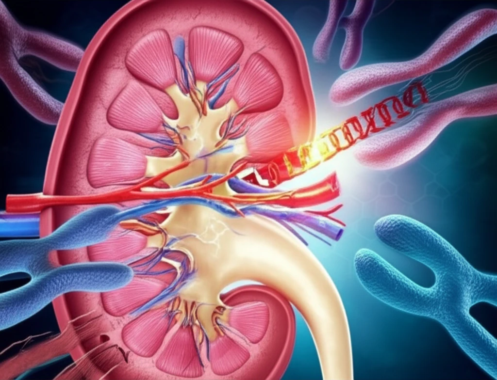
RCC with t(6;11)(p21;q12) is defined by a specific genetic abnormality: a translocation between chromosomes 6 and 11. This translocation results in the fusion of the Alpha gene to the TFEB gene. This fusion leads to the overexpression of the TFEB protein, which plays a significant role in the development and progression of this type of cancer.

While relatively rare, t(6;11) RCC has distinctive features that set it apart from other kidney cancers:

  • Age: Tends to occur in younger patients, often children and young adults, though cases in older adults have been reported.
  • Symptoms: Presentation can vary, from acute abdominal pain to incidental discovery during imaging for other reasons.
  • Macroscopic Appearance: Tumors often exhibit a grayish-white color on the cut surface, with potential for hemorrhage, cysts, and daughter nodules.
  • Microscopic Features: Characterized by two cell populations: large cells with clear cytoplasm and small cells forming rosette-like structures around basement membrane-like material.
  • Immunohistochemistry: Tumor cells consistently show positive staining for TFEB protein.
Accurate diagnosis is critical for appropriate management. Immunohistochemistry for TFEB and break-apart FISH (fluorescence in situ hybridization) for the TFEB gene are valuable tools for confirming the diagnosis. FISH analysis helps visualize the separation of the TFEB gene, confirming the translocation event.

Navigating the Challenges and Future Directions

While t(6;11) RCC generally has a favorable prognosis, some cases can exhibit aggressive behavior, including recurrence and metastasis. Therefore, ongoing monitoring and tailored treatment strategies are essential.

Targeted therapies, such as mTOR inhibitors, may be effective in certain cases, particularly those with metastatic disease. Immunotherapy and VEGF/PDGF inhibitors are also potential treatment options.

Continued research is crucial to better understand the underlying biology of t(6;11) RCC and to develop more effective therapies. Further studies are needed to identify predictive biomarkers and to refine treatment algorithms for this unique kidney cancer subtype. Awareness among urologists and pathologists, coupled with advanced diagnostic techniques, remains the cornerstone of improved outcomes for patients with t(6;11) RCC.

About this Article -

This article was crafted using a human-AI hybrid and collaborative approach. AI assisted our team with initial drafting, research insights, identifying key questions, and image generation. Our human editors guided topic selection, defined the angle, structured the content, ensured factual accuracy and relevance, refined the tone, and conducted thorough editing to deliver helpful, high-quality information.See our About page for more information.

This article is based on research published under:

DOI-LINK: 10.5114/pjp.2017.67617, Alternate LINK

Title: Clinicopathological Study Of 5 Cases Of Renal Cell Carcinoma With T(6;11)(P21;Q12)

Subject: General Medicine

Journal: Polish Journal of Pathology

Publisher: Termedia Sp. z.o.o.

Authors: Naoto Kuroda, Kenji Yorita, Naomi Sasaki, Akira Ishihara, Keiko Matsuura, Tsutomu Daa, Shintaro Mori, Aya Sasaki, Shuji Mikami, Kazuto Shigematsu, Yoji Nagashima

Published: 2017-01-01

Everything You Need To Know

1

What exactly defines renal cell carcinoma with t(6;11) translocation, and what makes it different from other kidney cancers?

Renal cell carcinoma with t(6;11) translocation is distinctive because it is defined by a specific genetic abnormality: a translocation between chromosomes 6 and 11, denoted as t(6;11)(p21;q12). This translocation results in the fusion of the Alpha gene to the TFEB gene, leading to the overexpression of the TFEB protein. While relatively rare, t(6;11) RCC has distinctive features that include the patient's age, symptoms, macroscopic appearance, microscopic features, and immunohistochemistry.

2

How is renal cell carcinoma with t(6;11) translocation accurately diagnosed, and what specific techniques are utilized in the diagnostic process?

Diagnosing renal cell carcinoma with t(6;11) translocation involves a combination of techniques. Immunohistochemistry for TFEB protein is a key diagnostic tool. Additionally, break-apart FISH (fluorescence in situ hybridization) for the TFEB gene is valuable for confirming the diagnosis, as FISH analysis helps visualize the separation of the TFEB gene, thereby confirming the translocation event.

3

What is the typical prognosis for renal cell carcinoma with t(6;11) translocation, and what factors influence its potential for aggressive behavior?

While renal cell carcinoma with t(6;11) translocation generally has a favorable prognosis, there are instances where it can exhibit aggressive behavior, including recurrence and metastasis. Therefore, ongoing monitoring and tailored treatment strategies are essential. The behavior underscores the importance of personalized treatment plans and vigilant follow-up care for individuals diagnosed with this rare kidney cancer subtype.

4

How does the t(6;11) translocation drive the development and progression, and how does the fusion of the Alpha gene to the TFEB gene affect cellular processes?

RCC with t(6;11)(p21;q12) translocation is defined by a specific genetic abnormality: a translocation between chromosomes 6 and 11. This translocation results in the fusion of the Alpha gene to the TFEB gene. This fusion leads to the overexpression of the TFEB protein, which plays a significant role in the development and progression of this type of cancer. Because the protein is overexpressed the cells have an increased ability to replicate.

5

Why is the World Health Organization (WHO) classification of RCC with t(6;11)(p21;q12) translocation as a distinct entity so significant in the context of kidney cancers?

The recent World Health Organization (WHO) classification of RCC with t(6;11)(p21;q12) translocation as a distinct entity is significant because it highlights the importance of recognizing this specific type of kidney cancer. This classification underscores the need for urologists and pathologists to be aware of its existence and unique features, so they can lead to earlier diagnosis and potentially more effective treatment strategies.

Newsletter Subscribe

Subscribe to get the latest articles and insights directly in your inbox.