Unlocking the Mysteries of Neuropathic Pain: How Functional Imaging Can Pave the Way to Better Treatments
"Explore how functional brain imaging is revolutionizing our understanding of neuropathic pain, offering new hope for chronic pain sufferers."
Neuropathic pain, a chronic condition resulting from nerve damage or dysfunction, affects millions worldwide. Unlike nociceptive pain, which arises from tissue injury, neuropathic pain stems from the nervous system itself, leading to sensations such as burning, shooting, or stabbing pain, often accompanied by tingling and numbness. This type of pain can be incredibly challenging to treat, as it doesn't respond well to conventional pain medications.
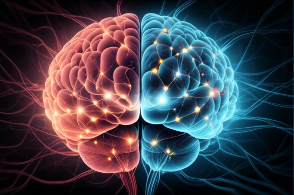
Functional brain imaging has emerged as a powerful tool in the quest to unravel the complexities of pain, particularly neuropathic pain. Since its application to pain research began in 1991, functional imaging has provided unprecedented insights into how the brain processes pain signals, identifies the specific brain regions involved, and reveals the changes that occur in chronic pain conditions. These techniques offer a window into the brain's activity, allowing researchers to map the neural circuits underlying pain perception and modulation.
This article explores how functional imaging techniques, such as fMRI (functional magnetic resonance imaging) and PET (positron emission tomography), are being used to study neuropathic pain. We will delve into the different types of studies conducted, the key brain regions implicated in pain processing, and the potential of these findings to lead to more effective and targeted treatments for neuropathic pain.
Decoding Brain Activity: The Four Main Types of Functional Imaging Studies in Pain Research
Functional imaging studies in pain research can be broadly categorized into four main types, each providing unique information about the brain's response to pain:
- Brain Responses to Noxious Stimuli: These studies compare brain activity during painful stimuli (e.g., heat) with activity during non-painful stimuli. The goal is to identify the brain regions specifically involved in processing pain signals.
- Identifies key brain structures involved in nociception.
- Serves as descriptive rather than explanatory.
- Lists brain structures that play a role in nociception.
- Empathy for Pain: These investigations explore brain activation when subjects observe or imagine others experiencing pain. This helps to differentiate between general emotional responses and pain-specific neural activity.
The Future of Pain Management: Functional Imaging as a Guide
Functional imaging holds immense promise for revolutionizing the diagnosis and treatment of neuropathic pain. By providing a deeper understanding of the neural mechanisms underlying chronic pain conditions, these techniques can pave the way for more targeted and personalized therapies. As research continues and technology advances, functional imaging is poised to become an indispensable tool in the fight against chronic pain, offering new hope for millions of sufferers worldwide.