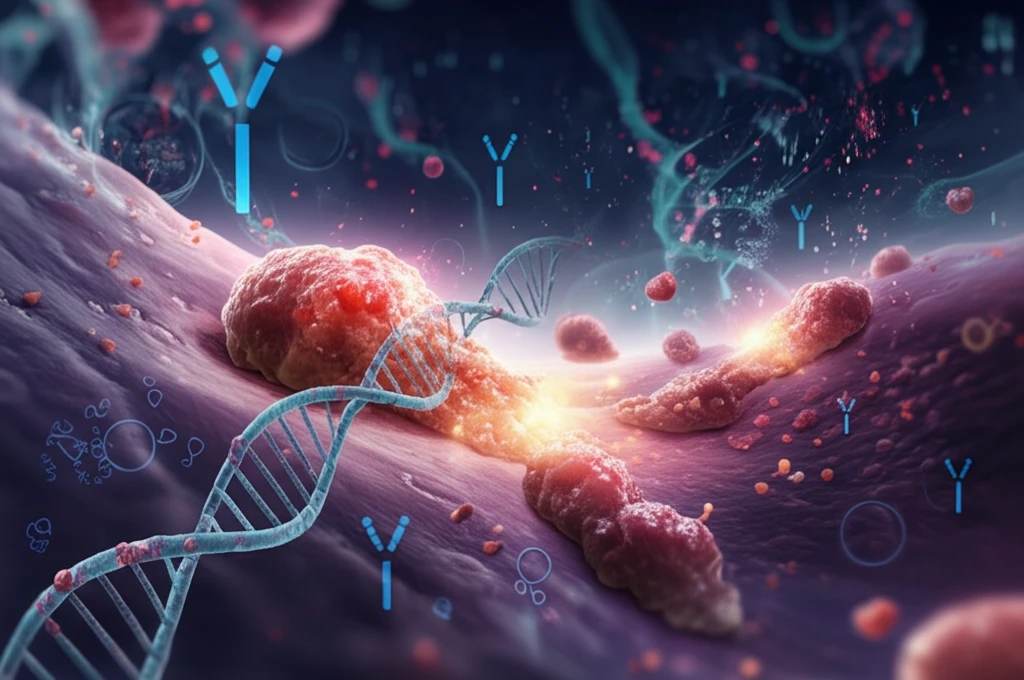
Microscopic view of liver cells transitioning to cancerous, DNA, biomarker.

Unlocking the Mysteries of Liver Cancer: A Comprehensive Guide to Histopathology and Diagnosis

"Navigating the complexities of hepatocellular carcinoma through histopathology, biomarkers, and the latest WHO classifications"


Hepatocellular carcinoma (HCC), a primary malignancy of the liver, stands as the sixth most prevalent cancer globally, marked by a concerningly high mortality rate and an escalating incidence. This malignancy's roots are often entwined with factors such as environmental exposures, dietary habits, and lifestyle choices. While HCC predominantly arises in livers already scarred by cirrhosis, a noteworthy shift has emerged: an increasing proportion of cases are now developing in livers with minimal or no fibrosis, signaling a change in the underlying causes and requiring updated diagnostic approaches.

The evolving landscape of HCC presents new challenges for pathologists. Traditional diagnostic criteria, while effective for progressed HCC, often fall short when dealing with small, early-stage lesions. These nascent tumors, frequently detected through routine screening programs, demand a more nuanced diagnostic approach. Adding complexity, recent reclassifications by the World Health Organization (WHO) have redefined lesions previously thought to originate from progenitor cells, necessitating a thorough understanding of these changes.

This article delves into the histopathology of HCC, summarizing recent advancements and contextualizing novel HCC biomarkers within the framework of the latest WHO reclassifications. Furthermore, it addresses the unique characteristics of combined hepatocellular-cholangiocellular carcinomas, offering a comprehensive overview of the diagnostic considerations for this complex malignancy.

Decoding HCC: From Precursor Lesions to Histologic Grading

Microscopic view of liver cells transitioning to cancerous, DNA, biomarker.

The journey of HCC often begins with precursor lesions, subtle changes within the liver that may or may not progress to full-blown cancer. Hepatocellular adenomas (HCAs), benign tumors of the liver, can, in rare instances, transform into HCC. These adenomas are more frequently observed in women using oral contraceptives, individuals with glycogen storage disease, or those undergoing androgen treatment. Metabolic syndrome has also emerged as a significant risk factor for HCA development. Differentiating between HCA and well-differentiated HCC, especially in non-cirrhotic livers, can be particularly challenging, requiring careful assessment of architectural and cytological features.

Dysplastic foci and nodules represent other critical precursor lesions. Dysplastic foci, small clusters of atypical cells, are characterized by uniform morphology and subtle deviations from surrounding liver tissue. Dysplastic nodules, larger than 1mm, are further categorized into low-grade and high-grade variants. While both are considered potential precursors to HCC, regression has also been observed. High-grade dysplastic nodules exhibit more pronounced cellular abnormalities, including increased nuclear size, irregular borders, and altered cellular arrangements.

  • Dysplastic Foci: Small clusters of atypical cells with uniform morphology.
  • Low-Grade Dysplastic Nodules: Minimal abnormalities with slight cellular changes.
  • High-Grade Dysplastic Nodules: More pronounced cellular abnormalities, indicating a higher risk of progression.
  • HCC: Definite malignant transformation with distinct invasive characteristics.
The gross pathology of HCC, or its appearance to the naked eye, varies depending on the stage and growth pattern of the tumor. Early-stage HCCs often present as vaguely nodular lesions, making them difficult to distinguish from surrounding cirrhotic tissue. Progressed HCCs, on the other hand, typically exhibit a distinctly nodular appearance, with a discernible capsule and potential for extracapsular growth. Macroscopic classifications include nodular, massive, and diffuse types, each reflecting different growth patterns and stages of the disease. Histologic grading, primarily using the Edmondson and Steiner system, further categorizes HCC based on cellular differentiation, nuclear irregularity, and pleomorphism, providing valuable prognostic information.

The Future of HCC Diagnostics

The ongoing refinement of diagnostic criteria, coupled with the discovery of novel biomarkers and a deeper understanding of the molecular underpinnings of HCC, promises to improve early detection, risk stratification, and personalized treatment strategies for this challenging malignancy. As research continues and technology advances, we can anticipate even more precise and effective approaches to combating liver cancer in the years to come.

About this Article -

This article was crafted using a human-AI hybrid and collaborative approach. AI assisted our team with initial drafting, research insights, identifying key questions, and image generation. Our human editors guided topic selection, defined the angle, structured the content, ensured factual accuracy and relevance, refined the tone, and conducted thorough editing to deliver helpful, high-quality information.See our About page for more information.

This article is based on research published under:

DOI-LINK: 10.3748/wjg.v20.i43.15955, Alternate LINK

Title: Histopathology Of Hepatocellular Carcinoma

Subject: Gastroenterology

Journal: World Journal of Gastroenterology

Publisher: Baishideng Publishing Group Inc.

Authors: Manuel Schlageter, Luigi Maria Terracciano, Salvatore D’Angelo, Paolo Sorrentino

Published: 2014-01-01

Everything You Need To Know

1

What is hepatocellular carcinoma and what are some of the challenges in diagnosing it?

Hepatocellular carcinoma, or HCC, is a primary liver malignancy and the sixth most common cancer globally. It often arises in livers scarred by cirrhosis, but is increasingly developing in livers with minimal or no fibrosis. The diagnosis of early-stage HCC can be challenging, requiring nuanced diagnostic approaches, especially with the recent WHO reclassifications of progenitor cell lesions. Combined hepatocellular-cholangiocellular carcinomas also present unique diagnostic considerations.

2

What are hepatocellular adenomas (HCAs) and what factors are associated with their development and potential transformation?

Hepatocellular adenomas, or HCAs, are benign liver tumors that, in rare cases, can transform into hepatocellular carcinoma. These adenomas are more common in women using oral contraceptives, individuals with glycogen storage disease, or those undergoing androgen treatment, with metabolic syndrome also recognized as a risk factor. The differentiation between HCA and well-differentiated HCC can be challenging, necessitating a careful evaluation of both architectural and cytological features.

3

What are dysplastic foci and nodules in the context of liver cancer, and how are they classified?

Dysplastic foci are small clusters of atypical cells characterized by uniform morphology, while dysplastic nodules are larger (greater than 1mm) and are categorized as low-grade or high-grade. Both are considered potential precursors to hepatocellular carcinoma, although regression has been observed. High-grade dysplastic nodules exhibit more pronounced cellular abnormalities, like increased nuclear size and irregular borders, indicating a higher risk of progression to HCC.

4

How does the macroscopic appearance and histologic grading of hepatocellular carcinoma inform diagnosis and prognosis?

The gross pathology of hepatocellular carcinoma varies by stage. Early-stage HCC often presents as vaguely nodular lesions, difficult to distinguish from surrounding cirrhotic tissue. Progressed HCCs typically exhibit a distinct nodular appearance, with a discernible capsule and potential for extracapsular growth. Macroscopic classifications include nodular, massive, and diffuse types. Histologic grading, notably using the Edmondson and Steiner system, categorizes HCC based on cellular differentiation, nuclear irregularity, and pleomorphism, providing valuable prognostic information. These methods help assess the cancer's progression and aggressiveness.

5

What advancements are expected to improve the future of hepatocellular carcinoma diagnostics, and what specific details are missing from this discussion?

The ongoing refinement of diagnostic criteria, discovery of novel biomarkers, and deeper understanding of the molecular underpinnings of hepatocellular carcinoma promise to improve early detection, risk stratification, and personalized treatment strategies. This includes better differentiation between benign and malignant lesions, prediction of disease progression, and tailoring therapies to individual patient profiles. However, the text does not detail specific biomarkers currently in use or under investigation, nor does it elaborate on specific personalized treatment strategies.

Newsletter Subscribe

Subscribe to get the latest articles and insights directly in your inbox.