A visually stunning depiction of a cell being analyzed with DNA-PAINT microscopy, showcasing the high-resolution imaging capabilities and the specific targeting of molecules within the cell. The image highlights the futuristic potential of this technology for biomedical research and diagnostics.

Unlocking the Microscopic World: How DNA-PAINT Revolutionizes Biomedical Research

"Dive into the groundbreaking technique of DNA-PAINT microscopy, transforming how we visualize cells and paving the way for new medical discoveries."


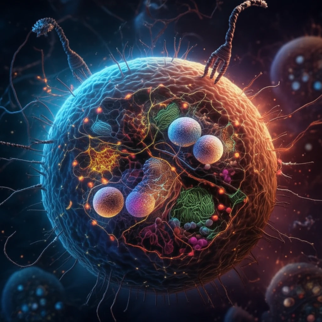
Imagine a world where you can zoom in on the tiniest parts of a cell, seeing details previously hidden from view. This is the promise of super-resolution microscopy, a revolutionary technology that's changing the landscape of biomedical research. Among the most exciting developments is DNA-PAINT (Points Accumulation for Imaging in Nanoscale Topography), a technique that allows scientists to visualize cellular structures with incredible precision.

Traditional microscopes have limitations, like the diffraction limit of light, which restricts how small an object can be distinguished. Super-resolution microscopy overcomes these limitations, enabling scientists to see details far beyond what was previously possible. This opens up new avenues for understanding how cells function, how diseases develop, and how we might intervene to treat them.

This article explores the innovative use of Affimers, small protein-based binding agents, combined with DNA-PAINT. This powerful combination offers a new approach to cellular imaging, with the potential to transform how we study cells and diagnose diseases. The integration of these techniques holds promise for more detailed insights into biological processes and, ultimately, better healthcare outcomes.

Deciphering DNA-PAINT: The Science of Visualizing at the Nanoscale

A visually stunning depiction of a cell being analyzed with DNA-PAINT microscopy, showcasing the high-resolution imaging capabilities and the specific targeting of molecules within the cell. The image highlights the futuristic potential of this technology for biomedical research and diagnostics.

DNA-PAINT works on a simple yet ingenious principle. It uses short strands of DNA, called 'docking strands', that are attached to specific molecules within a cell. These docking strands temporarily bind to complementary 'imager strands' that are labeled with fluorescent dyes. The transient binding and unbinding of the imager strands create a blinking effect, allowing scientists to pinpoint the location of the target molecules with remarkable accuracy.

This method has several advantages over traditional microscopy techniques. First, the blinking allows for high-resolution imaging. Second, the DNA strands are small, minimizing the interference with the natural structure of the cell. Finally, by using different DNA sequences, scientists can label and visualize multiple targets simultaneously, creating a detailed map of the cellular environment. DNA-PAINT's success relies on the transient binding of imager strands to docking strands.

  • High Resolution: DNA-PAINT surpasses the limitations of traditional light microscopy.
  • Specificity: The use of docking and imager strands ensures precise targeting of molecules.
  • Multiplexing: Multiple targets can be visualized simultaneously.
Affimers, which are small, engineered proteins that bind to specific target molecules, play a crucial role in this process. Researchers can attach DNA docking strands to Affimers, allowing them to specifically target and label molecules of interest within the cell. The use of Affimers, coupled with DNA-PAINT, offers a highly versatile and powerful approach to super-resolution microscopy.

The Future of Cellular Imaging: DNA-PAINT and Beyond

DNA-PAINT microscopy, combined with the specificity of Affimers, represents a major advancement in biomedical research. As technology continues to develop, we can expect even higher resolution, greater ease of use, and expanded applications. This innovative approach has the potential to unlock new discoveries and to provide more comprehensive understanding of human health and disease, opening doors to better diagnostics and more effective treatments. It is a testament to the power of innovation to push scientific boundaries.

About this Article -

This article was crafted using a human-AI hybrid and collaborative approach. AI assisted our team with initial drafting, research insights, identifying key questions, and image generation. Our human editors guided topic selection, defined the angle, structured the content, ensured factual accuracy and relevance, refined the tone, and conducted thorough editing to deliver helpful, high-quality information.See our About page for more information.

Everything You Need To Know

1

What is DNA-PAINT and why is it important in biomedical research?

DNA-PAINT (Points Accumulation for Imaging in Nanoscale Topography) is a super-resolution microscopy technique that allows scientists to visualize cellular structures with incredible precision, overcoming the limitations of traditional microscopes. Its importance lies in enabling researchers to see details far beyond what was previously possible, opening new avenues for understanding how cells function, how diseases develop, and how we might intervene to treat them. The method uses short strands of DNA, 'docking strands', attached to specific molecules within a cell. These docking strands temporarily bind to complementary 'imager strands' that are labeled with fluorescent dyes. The transient binding and unbinding of the imager strands create a blinking effect, allowing scientists to pinpoint the location of the target molecules with remarkable accuracy.

2

How does DNA-PAINT overcome the limitations of traditional light microscopy?

Traditional light microscopy is limited by the diffraction limit of light, restricting how small an object can be distinguished. DNA-PAINT overcomes this limitation through a process of transient binding of imager strands to docking strands, creating a blinking effect. This blinking allows scientists to pinpoint the location of target molecules with remarkable accuracy, achieving high-resolution imaging beyond the capabilities of traditional methods. Furthermore, the use of small DNA strands minimizes interference with the natural structure of the cell.

3

What are Affimers, and how are they used in conjunction with DNA-PAINT?

Affimers are small, engineered proteins that bind to specific target molecules. In the context of DNA-PAINT, researchers attach DNA docking strands to Affimers. This allows the Affimers to specifically target and label molecules of interest within the cell, enabling precise and versatile super-resolution microscopy. The combination of Affimers and DNA-PAINT offers a powerful approach to cellular imaging, enhancing the specificity of the technique.

4

What are the advantages of using DNA-PAINT for cellular imaging?

DNA-PAINT offers several key advantages for cellular imaging. Firstly, it provides high resolution, surpassing the limitations of traditional light microscopy. Secondly, the use of docking and imager strands ensures precise targeting of molecules. Thirdly, it allows for multiplexing, enabling the simultaneous visualization of multiple targets within the cell, creating a detailed map of the cellular environment. Furthermore, the DNA strands are small, minimizing interference with the cell's natural structure. These aspects combined allow a deeper understanding of cellular function. Missing from this approach is ease of use. As technology continues to develop, there is work to be done to make the application of DNA-PAINT more accessible.

5

What are the potential future applications of DNA-PAINT in biomedical research and healthcare?

DNA-PAINT microscopy, especially when combined with the specificity of Affimers, represents a major advancement with significant potential applications. Future developments promise even higher resolution, greater ease of use, and expanded applications in biomedical research. This innovative approach could unlock new discoveries and provide a more comprehensive understanding of human health and disease, potentially leading to better diagnostics, more effective treatments, and insights into biological processes. By being able to visualize multiple targets simultaneously DNA-PAINT can be applied to understanding complex disease processes.

Newsletter Subscribe

Subscribe to get the latest articles and insights directly in your inbox.