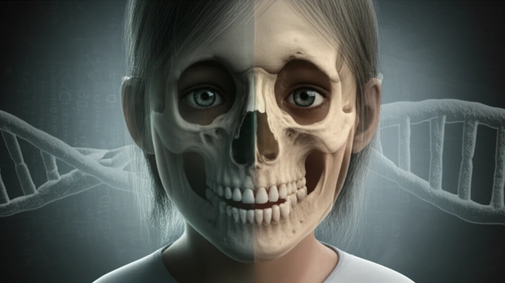
Unlocking the Clues: How a Dentist Can Spot Cleidocranial Dysostosis
"A Family's Journey to Diagnosis and the Importance of Early Detection"
Cleidocranial dysostosis (CCD) is a rare genetic disorder, affecting roughly 1 in 200,000 to 1 in 1,000,000 people. This condition primarily impacts the development of bones and teeth. It's characterized by a range of alterations, most notably in the clavicles, skull, face, and teeth, and practically affects the entire skeleton. CCD follows an autosomal dominant inheritance pattern, meaning it can be passed down from one parent to their child, regardless of sex or race.
The root cause of CCD lies in a defect in the CBFA1 gene, located on the 6p21 chromosome. This gene is pivotal for guiding the differentiation of osteoblast precursor cells, which are essential for the development of both endochondral and membranous bone tissues. The disruption in this gene's function can lead to delayed ossification—the process of bone formation—particularly in the skull, pelvis, and extremities.
Diagnosing CCD involves a careful evaluation of clinical signs and radiographic findings. A combination of multiple supernumerary (extra) teeth, partial or complete absence of the clavicles, and open sagittal suture and fontanelles (the soft spots on a baby's head) is considered a telltale sign of CCD. However, it's essential to rule out other conditions with similar features, such as pycnodysostosis, which is distinguished by fragile bones, short stature, and partial absence of hand and foot bones.
A Dentist's Discovery: Uncovering the Clues to CCD

In a recent case study, a 28-year-old male was referred to a geneticist after his dentist noticed supernumerary teeth and dysmorphic features. During the consultation, the patient reported a 'lump' in his left maxillary region following a tooth extraction. A physical examination revealed several notable characteristics: short stature, prominent cranial bones with incomplete fontanelle closure, subtle exophthalmos (protruding eyes), mid-face hypoplasia, an ogival palate, occlusal disharmony, and multiple carious lesions.
- Key Clinical Findings: Short stature, cranial bone abnormalities, dental anomalies.
- Radiographic Evidence: Supernumerary teeth, unerupted teeth, clavicle hypoplasia.
- Family History: Multiple relatives with similar characteristics.
Why Early Diagnosis Matters
This case highlights the critical role of dentists in identifying potential cases of CCD. The varied intensity and presentation of clinical manifestations underscore the importance of considering CCD in patients with dental anomalies and skeletal abnormalities. A thorough clinical examination, radiographic imaging, and detailed family history are essential for accurate diagnosis.
Early diagnosis allows for timely intervention and management of CCD-related complications. A multidisciplinary approach involving dentists, geneticists, and other specialists is crucial for providing comprehensive care. This includes addressing dental issues, monitoring skeletal development, and providing genetic counseling to affected individuals and their families.
By raising awareness among healthcare professionals and the public, we can improve early detection rates and ensure that individuals with CCD receive the support and care they need to optimize their health and quality of life. Remember, if you notice unusual dental or skeletal features in yourself or your family, consult a healthcare professional for further evaluation.
