Unlocking the Brain's Stress Response: How Key Circuits Impact Mental Well-being
"Research identifies specific brain neurons that, when activated by stress, may contribute to depression, offering new avenues for treatment."
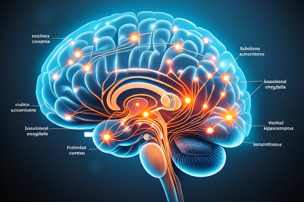
Stress is a common part of life, but for some, it can trigger more serious mental health issues like depression. The nucleus accumbens (NAc), a critical area in the brain, plays a significant role in how we respond to stress and experience related mood changes. This area is a key component of the ventral striatum, regulating mood and motivation.
The NAc doesn't work alone. It receives signals from other brain regions, including the ventral tegmental area (VTA), known for producing dopamine, a neurotransmitter linked to pleasure and reward. The NAc also gets input from areas like the prelimbic cortex (PL), basolateral amygdala (BLA), and ventral hippocampus (vHIP), all involved in processing emotions and memories. While the role of VTA-NAc neurons in stress response has been well-studied, the contribution of these other areas remains less clear.
Recent research has focused on understanding how these different brain regions contribute to the NAc's response to stress. One approach is to examine glutamatergic neurons, which use glutamate as a neurotransmitter and are known to play a role in activating other neurons. By studying these neurons, scientists hope to map out the brain circuits that become active under stress and how they influence the NAc.
Mapping the Brain's Response: Which Neurons Project to the Nucleus Accumbens Under Stress?
To investigate the glutamatergic pathways involved in the NAc's response to stress, researchers used a technique involving a retrograde tracer. This tracer, a fluorescent-tagged cholera toxin subunit B (CTB), was injected into the NAc of mice. CTB works by traveling backward along neurons, allowing scientists to identify which cells send signals into the NAc from other brain regions. After injecting the CTB, the mice were exposed to a stressor – in this case, restraint for one hour.
- Prelimbic Cortex (PL): The study found CTB-positive cells in the PL, indicating neurons projecting to the NAc. Further analysis revealed that 2.6% of these neurons were also activated by stress (co-labeled with c-Fos).
- Basolateral Amygdala (BLA): A similar pattern was observed in the BLA, with CTB-positive cells showing projections to the NAc. A higher percentage, 4.2%, of these neurons were also activated by stress.
- Ventral Hippocampus (vHIP): The vHIP also contained neurons projecting to the NAc, as indicated by CTB labeling. However, a smaller proportion, only 1.1%, of these neurons were activated by stress.
New Directions: Targeting Brain Circuits for Mental Wellness
This research provides valuable insights into the specific brain circuits involved in the stress response. By identifying the neurons in the PL, BLA, and vHIP that project to the NAc and are activated by stress, scientists are gaining a clearer picture of the complex pathways that contribute to mood disorders. This knowledge could pave the way for developing targeted interventions aimed at modulating the activity of these specific circuits and alleviating the symptoms of stress-related mental health conditions.