A detailed, artistic rendering of optoacoustic mesoscopy in action, visualizing skin layers, blood vessels, and the 'RSOM' technology at work.

Unlocking Skin Secrets: How a New Imaging Technique is Revolutionizing Dermatology

"Motion correction in optoacoustic mesoscopy is changing the way doctors diagnose and treat skin conditions."


Imagine a world where doctors can peer beneath the surface of your skin, not just to see what's visible, but to understand the intricate processes happening within. This is the promise of optoacoustic mesoscopy (RSOM), a revolutionary imaging technique that's changing the face of dermatology. By combining light and sound, RSOM offers a non-invasive way to visualize the skin's structure and detect signs of disease at a level of detail previously unattainable.

RSOM, also known as photoacoustic mesoscopy, is offering novel insights into vascular morphology and pathophysiological biomarkers of skin inflammation in vivo at depths unattainable by other optical imaging methods. However, motion effects may deteriorate performance and reduce the effective resolution. Now, researchers have developed a motion correction algorithm for RSOM.

This article delves into the fascinating world of RSOM, exploring its capabilities, the challenges it faces, and the groundbreaking advancements that are making it a powerful tool for dermatologists. We'll examine how this technology works, the impact it's having on patient care, and what the future holds for this exciting field.

What is Optoacoustic Mesoscopy and How Does it Work?

A detailed, artistic rendering of optoacoustic mesoscopy in action, visualizing skin layers, blood vessels, and the 'RSOM' technology at work.

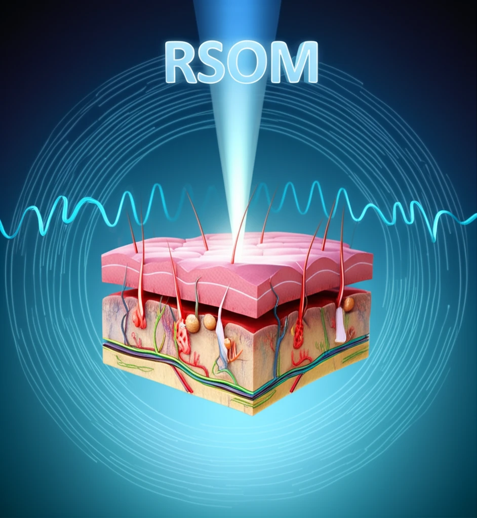
At its core, optoacoustic mesoscopy is a sophisticated imaging technique that uses pulses of light to generate sound waves within the skin. The light is absorbed by different components of the skin, such as blood vessels and melanin, causing them to expand and emit sound waves. These sound waves are then detected by an ultrasound transducer, which creates detailed images of the skin's internal structures.

RSOM image formation is achieved by raster-scanning a focused ultrasound detector over the region of interest (ROI) to collect optoacoustic waves generated in the tissue in response to pulsed laser illumination. The focal point of the ultrasound detector lies slightly above the surface of the sample, and the detector collects ultrasound signals over an acceptance angle (aperture) defined by the area and focusing characteristics of the detecting element. The collected data are tomographically reconstructed to yield an image of the light absorbers within the skin.

  • Non-Invasive: RSOM is a non-invasive technique, meaning it doesn't require any incisions or injections.
  • High Resolution: RSOM can achieve high-resolution images, allowing doctors to see details that are invisible to the naked eye.
  • Deep Penetration: RSOM can penetrate deeper into the skin than many other imaging techniques, providing a more complete picture of the skin's health.
  • Versatile: RSOM can be used to visualize a variety of skin conditions, from psoriasis to skin cancer.
The technology has recently demonstrated label free imaging and quantification of inflammatory biomarkers in psoriasis and eczema. Moreover, multi-wavelength illumination and spectral unmixing allows visualization of a larger number of pathophysiological features, including tissue/blood oxygenation, melanin patterns, or the bio-distribution of externally administered photo-absorbing labels.

The Future of Skin Imaging

The development of motion correction algorithms for RSOM is a significant step forward in the field of dermatology. As technology continues to advance, we can expect to see even more sophisticated imaging techniques that will enable doctors to diagnose and treat skin conditions with greater precision and effectiveness. With ongoing research, RSOM holds great promise for the future of skin health and well-being.

About this Article -

This article was crafted using a human-AI hybrid and collaborative approach. AI assisted our team with initial drafting, research insights, identifying key questions, and image generation. Our human editors guided topic selection, defined the angle, structured the content, ensured factual accuracy and relevance, refined the tone, and conducted thorough editing to deliver helpful, high-quality information.See our About page for more information.

Everything You Need To Know

1

What is optoacoustic mesoscopy (RSOM) and how does it provide insights into skin health?

Optoacoustic mesoscopy (RSOM) is an imaging technique that combines light and sound to visualize skin structures non-invasively. Pulses of light are absorbed by components like blood vessels and melanin, generating sound waves. These waves are detected to create detailed images, enabling the detection of diseases at a level of detail previously unattainable. It offers novel insights into vascular morphology and pathophysiological biomarkers of skin inflammation in vivo.

2

How does RSOM compare to other skin imaging methods in terms of invasiveness, resolution, and penetration depth?

RSOM stands out due to being non-invasive, meaning it doesn't require incisions or injections. It achieves high-resolution images, revealing details invisible to the naked eye. Additionally, RSOM can penetrate deeper into the skin than many other imaging techniques, offering a more complete view of skin health. This combination of features makes RSOM a versatile tool for visualizing various skin conditions.

3

What is the significance of motion correction algorithms in optoacoustic mesoscopy, and how do they improve image quality?

Motion correction algorithms are crucial for RSOM because motion effects can deteriorate performance and reduce the effective resolution of the images. By correcting for movement, these algorithms enhance the clarity and accuracy of RSOM images. The development of motion correction algorithms for RSOM is a significant step forward in the field of dermatology.

4

Beyond basic imaging, what advanced capabilities does optoacoustic mesoscopy offer for diagnosing and understanding skin conditions like psoriasis and eczema?

Optoacoustic mesoscopy allows for label-free imaging and quantification of inflammatory biomarkers in conditions like psoriasis and eczema. Multi-wavelength illumination and spectral unmixing can visualize a larger number of pathophysiological features, including tissue/blood oxygenation, melanin patterns, or the bio-distribution of externally administered photo-absorbing labels. While the technology shows great promise, the article did not mention how this technology can impact treatment.

5

What are the potential future advancements and implications of optoacoustic mesoscopy in dermatology, and how might it change patient care?

The future of optoacoustic mesoscopy in dermatology holds great promise with ongoing research and technological advancements. Future developments will enable doctors to diagnose and treat skin conditions with greater precision and effectiveness. It allows for unprecedented access to sub-dermal structures. The article did not mention cost factors associated with the equipment and the training. If such costs remain high, the technique might not be available to everyone.

Newsletter Subscribe

Subscribe to get the latest articles and insights directly in your inbox.