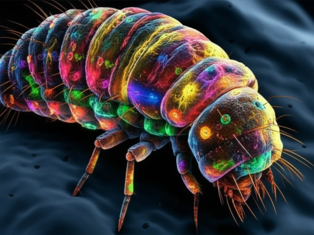
Drosophila larva with glowing neurons

Unlocking Neuron Secrets: A Beginner's Guide to Genetic Mosaics in Drosophila Research

"Dive into the innovative world of Drosophila larval sensory neuron analysis, exploring how genetic mosaics reveal critical insights into nervous system development and function."


The development of a complex nervous system hinges on precise neuron positioning and identity, followed by the development of unique dendritic structures and accurate axonal wiring. Dendritic Arborization (DA) sensory neurons in Drosophila larvae have become a leading model for deciphering the mechanisms of neuron differentiation.

With four primary classes of DA neurons, each exhibiting specific differences in dendritic complexity and genetic control, this system offers a practical way to study how neuron morphology is regulated. Researchers leverage the fruit fly's advanced genetic tools and the straightforward two-dimensional structure of the DA neuron dendrite, which is located just beneath the larval cuticle, to facilitate high-resolution in vivo visualization.

Diversity in dendritic morphology supports comparative analyses that identify key elements controlling the formation of simple versus complex dendritic trees. The stereotypical shapes of various DA neuron classes also allow for detailed morphometric statistical analyses. Understanding how these neurons function and connect can provide valuable insights into neural circuits and behavior.

Why Drosophila Larvae?

Drosophila larva with glowing neurons

Drosophila (fruit flies) provide an excellent model for studying genetics due to their short life cycle, ease of breeding, and well-characterized genome. Using Drosophila larvae, researchers can visually and genetically manipulate neurons to observe the development and function in ways that aren't possible in more complex organisms.

Genetic mosaics involve creating organisms with cells of different genotypes. This technique is used to study gene function in specific cells or tissues. Key methods in Drosophila research include:

  • MARCM (Mosaic Analysis with a Repressible Cell Marker): This technique allows researchers to selectively mark and manipulate specific cells, making it easier to study their development and function.
  • Flp-out: This method is used to induce gene expression or create mutant clones in specific cells at specific times, providing precise control over genetic modifications.
These methods allow scientists to create conditions where some neurons are genetically modified while others are not, providing a clear way to observe the effects of specific genes on neuronal development and behavior. By marking DA neurons through MARCM and Flp-out techniques, researchers gain valuable insights into neuronal development and function.

The Future of Neuron Research

The continued refinement of genetic tools and imaging techniques promises even more detailed insights into neuron development and function. By focusing on model organisms like Drosophila, researchers can uncover fundamental principles applicable across diverse species, contributing to our understanding of neurological disorders and potential therapeutic interventions.

About this Article -

This article was crafted using a human-AI hybrid and collaborative approach. AI assisted our team with initial drafting, research insights, identifying key questions, and image generation. Our human editors guided topic selection, defined the angle, structured the content, ensured factual accuracy and relevance, refined the tone, and conducted thorough editing to deliver helpful, high-quality information.See our About page for more information.

Everything You Need To Know

1

What are genetic mosaics, and how are they used in Drosophila research?

Genetic mosaics are organisms with cells of different genotypes. In Drosophila research, this technique is crucial for studying gene function in specific cells or tissues. By creating genetic mosaics, researchers can selectively manipulate certain neurons while leaving others unaffected, providing a clear way to observe the effects of specific genes on neuronal development and behavior, such as in the study of DA neurons.

2

Why is the study of Dendritic Arborization (DA) sensory neurons important, and what role do they play?

DA sensory neurons in Drosophila larvae are a leading model for understanding how neurons differentiate and develop, particularly regarding neuron specification, dendritic arborization, and axonal wiring. These neurons' stereotypical shapes and two-dimensional structure, located beneath the larval cuticle, make them ideal for high-resolution in vivo visualization and morphometric statistical analyses. The analysis of DA neurons helps in understanding neural circuits and behavior.

3

How do MARCM and Flp-out techniques contribute to the study of neuron development?

Both MARCM (Mosaic Analysis with a Repressible Cell Marker) and Flp-out techniques are essential tools in Drosophila research. MARCM allows researchers to selectively mark and manipulate specific cells, facilitating the study of their development and function. Flp-out induces gene expression or creates mutant clones in specific cells at specific times, providing precise control over genetic modifications. Together, these methods enable scientists to observe the effects of specific genes on neuronal development by creating conditions where some neurons are genetically modified while others are not.

4

What advantages does Drosophila offer as a model organism for studying neuron development, and why are Drosophila larvae used?

Drosophila, or fruit flies, provide an excellent model for studying genetics due to their short life cycle, ease of breeding, and a well-characterized genome. Using Drosophila larvae, researchers can visually and genetically manipulate neurons to observe development and function in ways not possible in more complex organisms. The larvae's DA neurons are particularly valuable due to their accessible location and straightforward two-dimensional structure.

5

What are the future prospects for neuron research using genetic mosaics and Drosophila, and what are the implications of these studies?

The continued refinement of genetic tools and imaging techniques promises even more detailed insights into neuron development and function. By focusing on model organisms like Drosophila, researchers can uncover fundamental principles applicable across diverse species, contributing to our understanding of neurological disorders and potential therapeutic interventions. Specifically, the study of DA neurons in Drosophila larvae offers valuable insights into neural circuits and behavior, with potential applications for human health.

Newsletter Subscribe

Subscribe to get the latest articles and insights directly in your inbox.